
Creative Biolabs is offering the most comprehensive services for antibody development projects. With strict regulation and effective execution, we are dedicated to providing the most valuable solutions to complete your projects.
HighlightsCreative Biolabs is offering the most comprehensive services for antibody development projects. With strict regulation and effective execution, we are dedicated to providing the most valuable solutions to complete your projects.
HighlightsCreative Biolabs has established a broad range of platforms for developing novel antibodies or equivalents. These cutting-edge technologies enable our scientists to meet your demands from different aspects and tailor the most appropriate solution that contributes to the success of your projects.
HighlightsWith deep understanding in antibody-related realms and extensive project experience, Creative Biolabs offers a variety of references to help you learn more about our capacities and achievements, including infographic, flyer, case study, peer-reviewed publications, and all kinds of knowledge that can assist your projects. You are also welcome to contact us directly for more specific solutions.
HighlightsGet a real taste of Creative Biolabs, one of the most professional custom service providers in the world. We are committed to providing highly customized comprehensive solutions with the best quality to advance your projects.
HighlightsScientists use M13 phage display as a versatile tool for multiple applications including protein interaction studies and the development of peptide-based drugs as well as antibodies. This technique uses the special characteristics of the M13 bacteriophage to attach peptides and proteins or other biomolecules to its exterior surface enabling researchers to build targeted libraries and perform affinity screening and molecular engineering. This article provides an extensive examination of the M13 phage display system by examining its structure and protocols while highlighting its applications and benefits compared to alternative display systems.
Researchers use phage display to link peptides and proteins onto bacteriophage surfaces which function as bacterial viruses. Phage display techniques utilize the M13 bacteriophage when they implement M13 phage display. The M13 bacteriophage's filamentous structure with single-stranded DNA and cylindrical form enables effective surface display. Genetic information within the M13 phage genome directs the production of proteins or peptides that are displayed on its surface
The M13 phage display system is widely used across several biological fields, including high-throughput screening of peptide libraries, antibody development, and protein engineering. Molecular biology, drug discovery processes and diagnostics now depend on this essential tool.
The M13 bacteriophage functions as a filamentous virus that targets Escherichia coli bacteria. The M13 bacteriophage extends to a length of 880 nanometers and keeps a diameter between 6 and 8 nanometers. The M13 phage coat consists of multiple copies of the major coat protein pVIII which arrange in a helical formation along its single-stranded DNA genome. The minor coat proteins pIII and pVII which are located at the extremities of the phage perform essential functions during interactions with host cells and in phage infection processes.
The M13 phage genome includes genetic instructions for structural proteins including pVIII, pIII, and pVI as well as replication components. By inserting foreign genes into the DNA encoding for pIII or pVIII, researchers can display peptides or proteins on the surface of the phage.
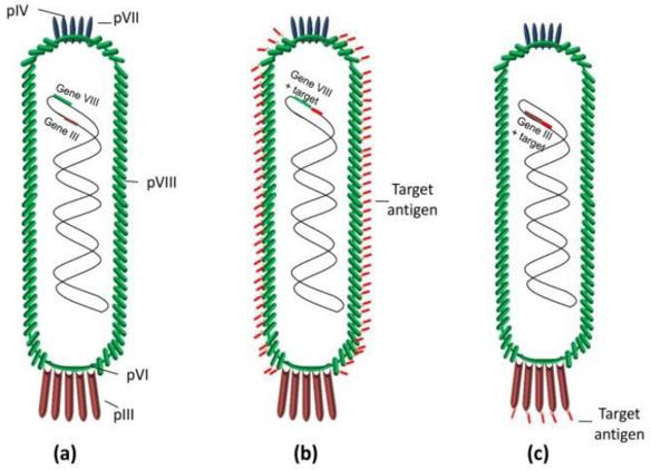 Fig. 1 Scheme of bacteriophage M13.1
Researchers commonly use the M13 bacteriophage in display systems due to its capability to infect E. coli cells as well as its surface display capacities for numerous proteins, peptides, and biomolecules. M13 phage display enables high-throughput screening and isolation of specific binding partners through surface-displayed recombinant proteins and peptides. M13 phage display serves as a perfect system for research in protein-protein interaction studies as well as peptide identification and antibody discovery while being useful in various biotechnological applications.
The process of amplifying M13 phage forms an essential part of phage display experiments. The fundamental steps to amplify M13 phage follow this procedure:
A phage display library is constructed by cloning DNA that encodes different peptides or proteins into the M13 phage genome. The library can be constructed using:
The constructed library enables phage particles to exhibit specific peptides and proteins on their surface resulting in a varied collection of surface-displayed biomolecules.
Phage display screening methods are used to isolate phages that specifically bind to a target molecule. Common techniques include:
The process of engineering M13 phage display requires consideration of multiple factors.
Scientists create cDNA libraries through a process that extracts mRNA from cells which they transform into complementary DNA before embedding these cDNA segments into the M13 phage. These libraries enable researchers to identify specific target bindings which serves as an important method for finding new proteins, antibodies, and ligands.
The M13 phage display system takes advantage of the distinct biological attributes of the M13 bacteriophage to present proteins or peptides on its exterior surface. The desired gene encoding a peptide or protein gets integrated into the M13 genome at the location of the minor coat protein (pIII) gene during this system's operation. Target protein or receptor interactions occur through displayed molecules which enable their selection and amplification.
Engineered M13 phage enters E. coli cells which replicate its genome and assemble new phage particles. The modified M13 phage displays recombinant proteins or peptides on its surface to facilitate specific target molecule interactions. Researchers can choose strong binders from phage particles that demonstrate superior binding affinity during the screening process.
Vaccine development relies heavily on the M13 phage display system which presents viral or bacterial antigens to trigger immune responses. Phage-displayed antigens function as essential tools to identify vaccine candidates and create vaccines against infectious diseases.
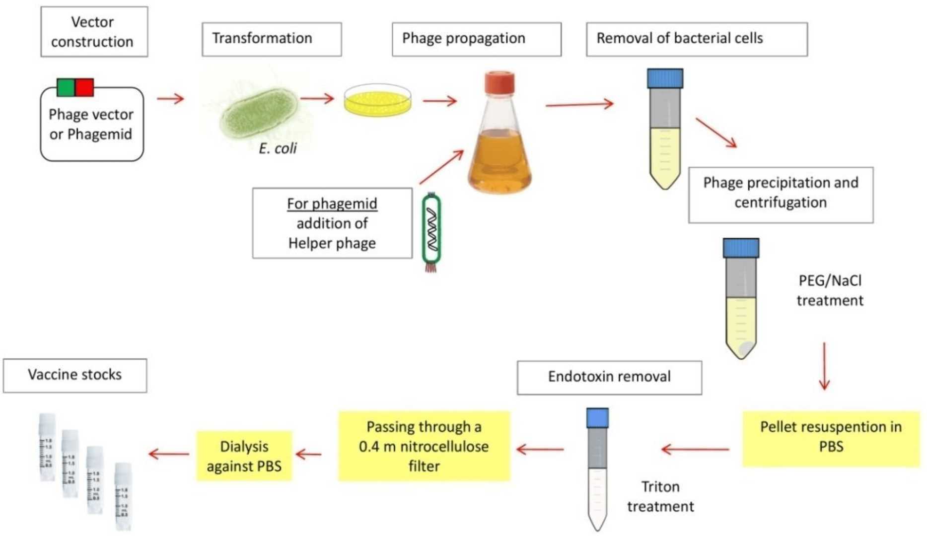 Fig. 2 Schematic of the preparation for M13 phage-based vaccine.1
The phage display technique stands out as a superior method for investigating protein-protein interactions. Researchers use M13 surface-displayed peptides or proteins to determine binding partners which helps to map out complex signaling networks.
M13 phage display continues to serve as a basic technique for antibody discovery. Researchers utilize phage display technology to display peptide libraries or antigen fragments on surfaces in order to Isolate antibodies which demonstrate strong specificity and high binding affinity for their targets.
Through the use of phage display technology scientists can discover peptides that demonstrate therapeutic efficacy. Through the screening of phage-displayed peptide libraries researchers discover peptides with binding affinity to disease-related targets leading to new drug candidates.
Scientists have used the M13 phage display system to create nanomaterials including phage-based nanoparticles which serve drug delivery purposes. The development of biosensors and diagnostic tools depends heavily on phage display technology.
Diagnostic tools development utilizes phage display to create assays that detect pathogens and biomarkers. Diagnostic kits utilize phage-displayed antibodies and peptides for improved sensitivity and specificity.
All listed services and products are For Research Use Only. Do Not use in any diagnostic or therapeutic applications.








