
Creative Biolabs is offering the most comprehensive services for antibody development projects. With strict regulation and effective execution, we are dedicated to providing the most valuable solutions to complete your projects.
HighlightsCreative Biolabs is offering the most comprehensive services for antibody development projects. With strict regulation and effective execution, we are dedicated to providing the most valuable solutions to complete your projects.
HighlightsCreative Biolabs has established a broad range of platforms for developing novel antibodies or equivalents. These cutting-edge technologies enable our scientists to meet your demands from different aspects and tailor the most appropriate solution that contributes to the success of your projects.
HighlightsWith deep understanding in antibody-related realms and extensive project experience, Creative Biolabs offers a variety of references to help you learn more about our capacities and achievements, including infographic, flyer, case study, peer-reviewed publications, and all kinds of knowledge that can assist your projects. You are also welcome to contact us directly for more specific solutions.
HighlightsGet a real taste of Creative Biolabs, one of the most professional custom service providers in the world. We are committed to providing highly customized comprehensive solutions with the best quality to advance your projects.
HighlightsPhage display is a powerful molecular biology tool that allows studying protein-protein interactions, identifying new peptides and selecting high-affinity antibodies. In widespread use in drug design, protein engineering and therapeutic antibody discovery, phage display is an ideal way to screen libraries of peptides, antibodies and proteins of any sort. Here we provide the phage display protocol, step by step instructions and troubleshooting.
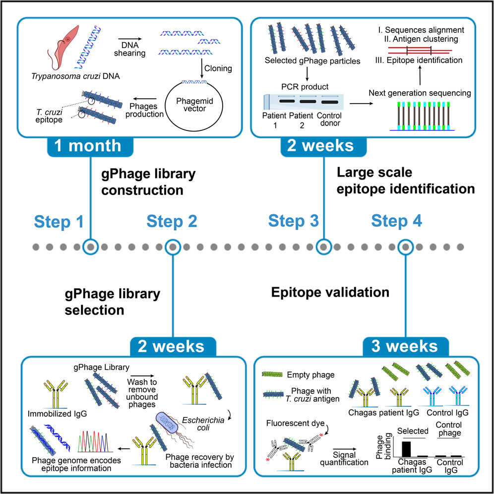 Fig. 1 Graphical abstract of protocol for design, construction, and selection of genome phage (gPhage) display libraries.1
Before beginning a phage display experiment, it is essential to gather the necessary materials and reagents. Proper preparation ensures a smoother process and more reliable results.
Phage display experiments require a variety of laboratory equipment, some of which are standard items in molecular biology labs. The following is a list of commonly used tools.
Table 1. Commonly used equipment in phage display experiment
| Equipment | Purpose |
| Microcentrifuge | To separate phage particles from other materials. |
| Magnetic beads | For capturing antigen-bound phages in biopanning. |
| Incubator | To grow bacterial cultures and phages. |
| Plate reader | To quantify absorbance in ELISA assays. |
| PCR machine | To amplify phage DNA for cloning and screening. |
| Refrigerated centrifuge | For pelleting cells or phages at low temperatures. |
| Shaking incubator or orbital shaker | For optimal bacterial growth and phage propagation. |
| Flow cytometer | For advanced analysis of phage binding profiles. |
| Gel electrophoresis equipment | For verifying phage DNA integrity and cloning success. |
| Phage ELISA setup | To screen for antigen-binding phages. |
The following are key reagents and supplies essential for conducting phage display:
Phage Vectors
Antigens and Targets for Screening
Buffer and Solution Recipes
Miscellaneous Materials
| Simple Antibody Phage Display | Phage Display for Peptide Libraries | Phage Display for Protein-Protein Interactions | |
| Description | Utilize phage vectors to display scFvs or Fab fragments on the phage surface. | Display random peptide sequences on the phage surface. Peptides bind specifically to the target. | Use phage display to identify binders for a target protein by exposing the target to phage display proteins. |
| Goal | To isolate monoclonal antibodies that bind specifically to the antigen. | To identify peptides that bind tightly to a target protein or cell. | To discover proteins that interact with a target protein. |
| Process | The library of antibodies is exposed to the antigen, and phages binding to the antigen are enriched through biopanning. | Peptide libraries are displayed on phages and subjected to biopanning against immobilized antigens. | The protein of interest is immobilized, and phage libraries of other proteins are screened for binding interactions. |
| Application |
Identifying monoclonal antibodies against specific antigens. Generating therapeutic antibodies. |
Screening for peptides with high affinity and specificity for proteins, cells, or small molecules. |
Mapping protein-protein interactions. Discovering new protein complexes and signaling pathways. |
The first step is to create a diverse library. The libraries can contain peptides, antibodies, or proteins.
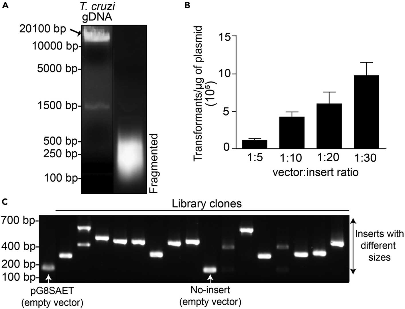 Fig. 2 Building and quality control of a Trypanosoma cruzi genomic DNA library.1
After biopanning, the selected phages are amplified to increase their concentration and enrich for high-affinity binders.
Once the enrichment process has been completed, the positive clones are analyzed.
All listed services and products are For Research Use Only. Do Not use in any diagnostic or therapeutic applications.








