
Creative Biolabs is offering the most comprehensive services for antibody development projects. With strict regulation and effective execution, we are dedicated to providing the most valuable solutions to complete your projects.
HighlightsCreative Biolabs is offering the most comprehensive services for antibody development projects. With strict regulation and effective execution, we are dedicated to providing the most valuable solutions to complete your projects.
HighlightsCreative Biolabs has established a broad range of platforms for developing novel antibodies or equivalents. These cutting-edge technologies enable our scientists to meet your demands from different aspects and tailor the most appropriate solution that contributes to the success of your projects.
HighlightsWith deep understanding in antibody-related realms and extensive project experience, Creative Biolabs offers a variety of references to help you learn more about our capacities and achievements, including infographic, flyer, case study, peer-reviewed publications, and all kinds of knowledge that can assist your projects. You are also welcome to contact us directly for more specific solutions.
HighlightsGet a real taste of Creative Biolabs, one of the most professional custom service providers in the world. We are committed to providing highly customized comprehensive solutions with the best quality to advance your projects.
HighlightsAt their core, proteins are linear polymers constructed from amino acids. There are typically 20 common types of amino acids found in proteins, each possessing a central carbon atom, an amino group, a carboxyl group, a hydrogen atom, and a variable side chain (R-group). The distinct arrangement and chemical characteristics of the R-groups determine both the structure and functionality of the protein. Amino acids link together via peptide bonds to form long chains known as polypeptides. A functional protein may consist of one or several polypeptide chains.
The saying "structure dictates function" proves most accurate within the field of protein science. The biological activity of a protein depends on the specific three-dimensional arrangement of its atoms. This includes:
Drug discovery along with protein engineering and disease mechanism research depend heavily on comprehensive knowledge of protein structures.
To simplify the complexity, protein structure is conventionally described at four hierarchical levels:
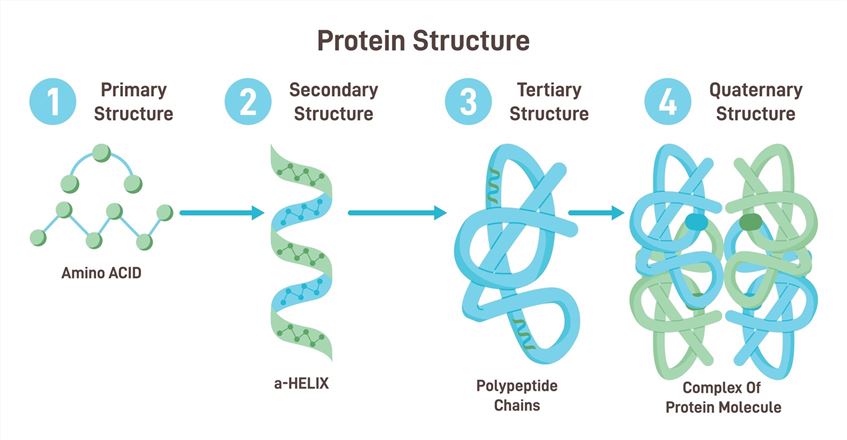 Fig. 1 Protein structure.
The primary structure represents a sequential arrangement of amino acids extending from the N-terminus to the C-terminus throughout a polypeptide chain. The primary structure functions identically to the arrangement of letters forming a word.
Genetic information establishes the primary structure of proteins. The gene's DNA nucleotide sequence directs messenger RNA (mRNA) formation followed by ribosomal translation into a precise amino acid sequence during protein creation.
Peptide bonds form the link between amino acids in protein structures. The peptide bond represents an amide bond created between the carboxyl group (−COOH) of one amino acid and the amino group (−NH2) of an adjacent amino acid through a condensation reaction that releases a water molecule.
Key characteristics of the peptide bond:
The primary structure is the fundamental level. The protein's higher-level structures (secondary through quaternary) can be defined from its intrinsic information under specific physiological conditions. One mutation in the primary sequence can produce significant changes to the 3D structure which may eliminate or change the protein's function.
The secondary structure designates the repeating structural patterns found among neighboring amino acids within a polypeptide chain. The secondary structure emerges through hydrogen bonding between peptide bond backbone amide hydrogens (−NH) and carbonyl oxygens (−C=O) while side chains remain uninvolved.
Two major types of secondary structure are particularly stable and widespread: alpha helix (α-helix) and beta pleated sheet (β-sheet).
Table 1. Comparison of Common Secondary Structures
| Feature | Alpha Helix (α-helix) | Beta Pleated Sheet (β-sheet) |
| Shape | Right-handed coil / Rod-like | Extended sheet / Zig-zag backbone |
| H-Bonding | Intra-chain | Inter-strand (Between adjacent strands) |
| Residues/Turn | ~3.6 | ~2 (per strand dimension) |
| R-Group | Project outwards from helix axis | Project alternately above/below sheet |
| Common in | Globular proteins, transmembrane domains, keratin | Globular proteins, silk fibroin |
The primary stabilization force for secondary structures stems from hydrogen bonds between the backbone peptide groups.
The tertiary structure defines how a complete polypeptide chain folds in three dimensions by positioning its secondary structures and amino acid side chains (R-groups). This aspect establishes whether the protein appears as a globular form or a fibrous structure.
The tertiary structure remains stable due to interactions involving amino acid side chains (R-groups) as well as connections between these side chains and the backbone.
The specific tertiary structure creates functional sites, such as:
Table 2. Interactions Stabilizing Tertiary Structure
| Interaction Type | Description | Relative Strength | Residues Involved (Examples) |
| Hydrophobic Interaction | Clustering of nonpolar side chains away from water | Moderate (cumulative) | Val, Leu, Ile, Met, Phe, Trp, Ala |
| Hydrogen Bond | Sharing of H atom between electronegative atoms (N, O) | Weak to Moderate | Ser, Thr, Tyr, Asn, Gln, His, Backbone |
| Ionic Bond (Salt Bridge) | Electrostatic attraction between opposite charges | Moderate to Strong | Lys(+), Arg(+) <-> Asp(-), Glu(-) |
| Disulfide Bridge | Covalent bond between two Cysteine sulfur atoms | Strong (Covalent) | Cys <-> Cys |
| Van der Waals Forces | Weak, short-range attractions due to fluctuating electron clouds | Weak (cumulative) | All atoms in close proximity |
The quaternary structure describes how several polypeptide chains or subunits arrange themselves spatially to form proteins with multiple chains called oligomeric proteins. Proteins made up of a single polypeptide chain lack quaternary structure.
Proteins that consist of multiple polypeptide chains exhibit this structural level. Examples include:
The same types of interactions that stabilize tertiary structure hold subunits together in quaternary structure at their interfaces.
The precise organization of subunits determines the effectiveness of protein regulation and functionality.
Protein folding describes how a polypeptide chain transforms into its functional three-dimensional native structure. Protein folding processes operate on the primary sequence and remain highly complex.
Proper protein folding is vital since misfolded proteins become non-functional and play a role in multiple diseases such as Alzheimer's and Parkinson's.
Denaturation is the loss of the native secondary, tertiary, and quaternary structure of a protein, leading to loss of function. The primary structure (sequence) remains intact. Common denaturing agents include:
The removal of denaturing agents sometimes allows reversibility of denaturation but extended exposure to harsh conditions makes it irreversible.
Determining the primary structure is fundamental:
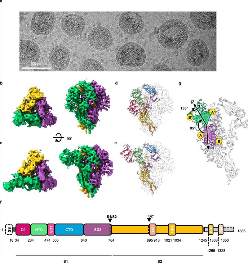 Fig. 2 In situ cryo-EM analysis of the structure of PEDV PT52 S on intact viruses.1
Protein structure is a vast and intricate field, fundamental to understanding nearly all biological processes. The ability to determine, predict, and analyze protein using experimental techniques like X-ray crystallography, NMR, Cryo-EM, combined with de novo sequencing platform, fuels innovation in medicine and biotechnology. At Creative Biolabs, we provide customized services for de novo sequencing and protein structural analysis to our customers.
Learn more about Creative Biolabs' de novo antibody sequencing services:
All listed services and products are For Research Use Only. Do Not use in any diagnostic or therapeutic applications.








