
Creative Biolabs is offering the most comprehensive services for antibody development projects. With strict regulation and effective execution, we are dedicated to providing the most valuable solutions to complete your projects.
HighlightsCreative Biolabs is offering the most comprehensive services for antibody development projects. With strict regulation and effective execution, we are dedicated to providing the most valuable solutions to complete your projects.
HighlightsCreative Biolabs has established a broad range of platforms for developing novel antibodies or equivalents. These cutting-edge technologies enable our scientists to meet your demands from different aspects and tailor the most appropriate solution that contributes to the success of your projects.
HighlightsWith deep understanding in antibody-related realms and extensive project experience, Creative Biolabs offers a variety of references to help you learn more about our capacities and achievements, including infographic, flyer, case study, peer-reviewed publications, and all kinds of knowledge that can assist your projects. You are also welcome to contact us directly for more specific solutions.
HighlightsGet a real taste of Creative Biolabs, one of the most professional custom service providers in the world. We are committed to providing highly customized comprehensive solutions with the best quality to advance your projects.
HighlightsThe precision of molecular recognition within the body is governed by a sophisticated landscape of cell surface receptors, extracellular matrix components, and tissue specific microenvironments. While traditional in vitro selection methods provide a controlled setting for identifying high affinity binders, they often fail to account for the structural complexity and physiological relevance of native biological systems. Conversely, in vivo screening, while biologically authentic, presents significant logistical and ethical challenges. Ex vivo phage display screening emerges as a critical intermediary methodology. By utilizing freshly isolated tissues or primary cell populations maintained in an external laboratory environment, this technique allows researchers to interrogate the molecular topography of living systems with high resolution and experimental accessibility.
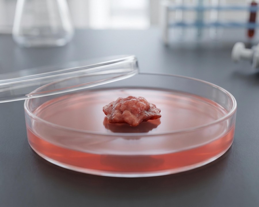
Ex vivo screening refers to the biopanning of phage display libraries, including peptides, scFv, Fab, or single domain antibodies, against biological materials that have been removed from an organism but retain their architectural and functional integrity. This approach bridges the gap between the simplicity of recombinant protein assays and the multifaceted nature of whole animal models. The process involves the direct incubation of a diverse genetic repertoire with target materials such as fresh frozen tissue sections, perfused organs, tumor biopsies, or patient derived xenografts.
The fundamental objective of this strategy is to identify ligands that recognize antigens in their natural conformation. Unlike purified proteins that are often immobilized on synthetic surfaces, targets in an outside the organism setting remain embedded within their native lipid bilayers or extracellular scaffolds. This preservation is vital for membrane proteins, particularly multi pass transmembrane receptors like G protein coupled receptors (GPCRs), which often undergo denaturation or misfolding when isolated from their cellular context. By presenting the library to an intact tissue architecture, researchers can isolate clones that bind to biologically meaningful epitopes that might be hidden or absent in simplified biochemical assays.
The decision to utilize in isolated tissues selection is typically driven by the need for physiological relevance when in vivo studies are restricted or when in vitro models are insufficient. Several specific research contexts benefit from this approach:
Ethical and safety constraints often prevent the systemic administration of phage libraries into human subjects. In isolated tissues platforms allow the use of ethically procured human tissues, such as surgical biopsies or organ donor material, to identify human specific targeting ligands for clinical applications.
Malignant tissues are characterized by a unique stromal architecture and heterogeneous cell populations. Ex vivo screening on fresh tumor sections or explanted cell sheets enables the discovery of binders that can penetrate the dense tumor matrix and recognize cancer specific markers in their native state.
Diseases involving significant tissue remodeling, such as liver cirrhosis or pulmonary fibrosis, create complex structural changes. Screening against these diseased tissue explants allows for the identification of ligands that home to specific pathological signatures.
In isolated tissues models of vascular endothelium or brain slices maintain the specialized junctional proteins and transporters required for studying tissue penetration and receptor mediated transport.
Ex vivo phage display offers a unique set of technical benefits that prioritize both the quality of the identified binders and the efficiency of the discovery process.
By using primary tissues or intact cells, the antigen density and spatial orientation remain consistent with the biological reality. This ensures that the identified antibodies or peptides have a higher probability of success when transitioned back into a living system.
Unlike in vivo screening, where systemic circulation and rapid renal clearance can limit the interaction time, in isolated tissues assays allow for precise control over incubation temperature, buffer composition, and washing stringency. This precision reduces the noise typically associated with animal models.
Advanced ex vivo protocols incorporate pre depletion steps, where the library is first incubated with nontarget tissues or cells. This negative selection removes common binders, ensuring that the remaining population is enriched for clones with true specificity for the target site.
Isolated tissues samples are compatible with a variety of sophisticated recovery techniques. For labeled target cells, researchers can utilize cell sorting to isolate the specific phage bound population with high purity.
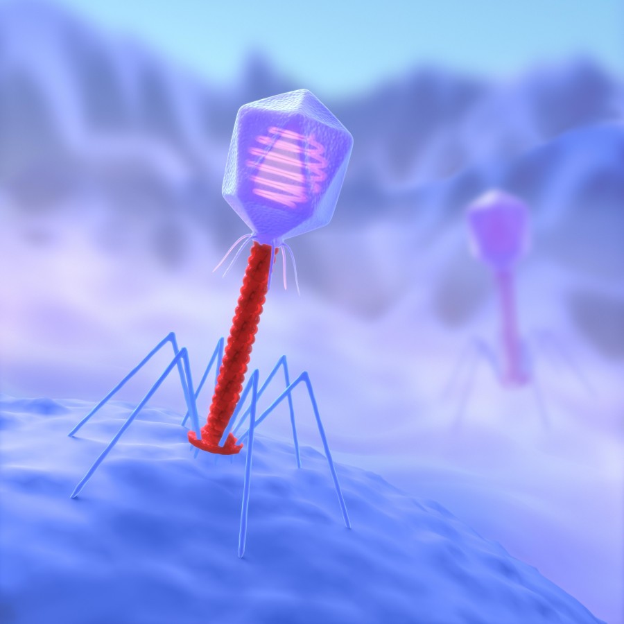
Creative Biolabs has established a high throughput Phage Display Platform specifically designed to handle the nuances of ex vivo selection. Our expertise ensures that biological materials are processed with minimal disruption to their structural integrity, maximizing the chances of recovering rare, high affinity clones. To support the global research community, we offer a specialized suite of services centered on bacteriophage display based binder discovery.
| Services | Our Capabilities |
| Bacteriophage Display Library Construction Service | We design and build high diversity libraries in scFv, Fab, or peptide formats, optimized for stability in diverse biological buffers. |
| Phage Display Library Screening Service | Our team provides tailored biopanning strategies against human biopsies, animal disease models, and specialized cell populations. |
| Monoclonal Antibody Discovery developed from Bacteriophage Display | A pipeline to isolate and characterize therapeutic grade antibodies with validated tissue specificity |
| Peptide Discovery Service by Bacteriophage Display | Identifying short, bioactive peptides for use in drug delivery, imaging, or as receptor modulators. |
| Stable Binder Discovery grounded in Phage Display | Engineering binders with enhanced biophysical properties to ensure performance in demanding physiological conditions. |
| pH Sensitive Binder Discovery based on Bacteriophage Display | Developing recycling antibodies that bind or release their targets in response to environmental pH shifts. |
| Internalizing Antibody Discovery developed from Phage Display | Specialized screening for ligands that are not only tissue specific but also capable of receptor mediated endocytosis. |
All listed services and products are For Research Use Only. Do Not use in any diagnostic or therapeutic applications.








