"Creative Biolabs is committed to providing highly customized comprehensive solutions with the best quality to advance our global clients’ projects."
In Vivo Phage Library Screening Service for Human Synovium Targeting
Background Platform Workflow Advantage Related Service FAQ Online Inquiry
The quest for site-specific therapeutic delivery remains the "Holy Grail" of modern pharmacology, particularly within the complex microenvironment of inflammatory joint diseases. Creative Biolabs, a global leader in phage display technology with over two decades of specialized experience, provides a premier Human Synovium Targeting In Vivo Phage Library Screening Service. Our platform is designed to identify high-affinity, tissue-specific ligands that can navigate the systemic circulation and home specifically to the human synovium. By bypassing the limitations of traditional in vitro panning, our in vivo strategies ensure that the identified candidates possess the necessary pharmacokinetic and biodistribution profiles to function in a living physiological system.
Discuss Your Project Today
The Strategic Importance of Human Synovium Targeting
The human synovium is a specialized connective tissue that lines the inner surface of joint capsules. In healthy individuals, it maintains joint homeostasis; however, in autoimmune conditions like Rheumatoid Arthritis (RA), the synovium undergoes massive hyperplasia, inflammatory infiltration, and neoangiogenesis. Traditional systemic administration of anti-inflammatory drugs often leads to off-target effects and suboptimal local concentrations. Human Synovium Targeting In Vivo Phage Library Screening addresses this by identifying peptides or antibody fragments (scFv/Fab) that specifically recognize synovial vascular markers or stromal components. This precision targeting enables:
-
Selective Drug Delivery: Conjugating therapeutic payloads (cytostatics, biologics, or siRNA) to synovial-homing peptides.
-
Molecular Imaging: Developing targeted contrast agents for early-stage diagnosis and monitoring of joint inflammation.
-
Gene Therapy: Engineering viral vectors with synovial-homing motifs for localized gene expression.
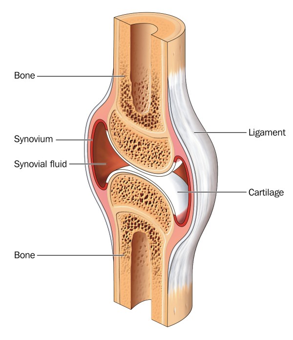 Fig. 1 Human synovium.
State-of-the-Art In Vivo Platform: Human Synovium Xenograft & Humanized Joint Models
At the heart of our service lies the sophisticated integration of human pathology into a living host system. Creative Biolabs has perfected the use of humanized models to bypass the "species gap" that often causes preclinical candidates to fail in human trials.
Human Synovium Xenograft Model
This model is the gold standard for translational rheumatology. It involves the surgical transplantation of fresh, viable human synovial tissue (obtained from synovectomies or arthroplasty) into immunodeficient mice.
-
Histological Fidelity: The xenograft preserves the original 3D architecture of the human synovium, including the distinct lining and sublining layers, as well as the endogenous human stromal and immune cell populations.
-
Vascular Integration: Over a period of several weeks, the host mouse's circulatory system undergoes anastomosis with the human synovial vessels. When a phage library is injected intravenously, the phages are carried through this chimeric circulation, requiring them to recognize human-specific vascular addressins to home to the graft.
-
Dynamic Microenvironment: The model captures the complex interplay between the extracellular matrix (ECM), interstitial pressure, and localized cytokine gradients (e.g., TNF-α, IL-1, IL-6, and IL-17) that define the inflammatory niche.
Humanized Mouse Joint Models
For broader applications, we employ Humanized Mouse Joint Models that simulate the holistic environment of a human joint.
-
Target-Humanized Mice: Utilizing genetically engineered mouse strains that express human versions of specific synovial targets, such as humanized TNF-α or humanized integrin receptors.
-
Cell-Based Humanization: Intra-articular implantation of primary human synovial fibroblasts (FLS) or macrophages into the joint space to study cell-specific homing and tissue-invasive behavior.
Advantages Over Conventional Screening
-
Physiological Selection: Phages must survive blood proteases, escape hepatic and splenic sequestration, and actively extravasate into the synovium.
-
Conformational Accuracy: Unlike in vitro panning on recombinant proteins, in vivo screening ensures ligands recognize targets in their native, multi-protein complex states within a living tissue.
Comprehensive Workflow: From Library Construction to Lead Validation
Creative Biolabs provides a seamless, end-to-end pipeline for Human Synovium Targeting In Vivo Phage Library Screening.
 Phase I: Customized Phage Library Design
We offer a vast array of high-diversity proprietary libraries (>1013 cfu), including:
-
Linear & Cyclic Peptide Libraries (7-mer, 12-mer, C7C): Ideal for identifying small, stable homing motifs.
-
Human scFv/Fab Libraries: Direct discovery of fully human antibody fragments.
-
VHH (single domain antibody) Libraries: Small, robust single-domain antibodies with superior tissue penetration.
Phase II: Model Preparation
Our team procures high-quality human synovial tissue through established clinical partnerships. Following rigorous quality control and histological verification, the tissue is grafted. We monitor graft viability and vascularization using non-invasive imaging before initiating the biopanning process.
Phase III: In Vivo Biopanning and Enrichment
-
Injection & Circulation: The library is administered systemically. We optimize circulation times (typically 30–90 minutes) based on the library type.
-
Systemic Perfusion: To eliminate non-specific background, the animal is perfused with heparinized saline, ensuring only high-affinity, tissue-bound phages remain.
-
Tissue Recovery: The human xenograft and control mouse organs (liver, lung, heart) are harvested.
-
Phage Elution: Bound phages are recovered through competitive elution or bacterial infection.
-
Iterative Selection: Usually, 3 to 5 rounds are performed to achieve logarithmic enrichment of synovial-specific clones.
Phase IV: Next-Generation Sequencing (NGS) and Bioinformatics
We utilize NGS-based Phage Display Analysis to provide a "big data" view of the selection process.
-
Clonal Evolution Tracking: Monitoring how specific motifs enrich across rounds.
-
Sequence Clustering: Identifying consensus motifs that indicate a common binding site.
-
Filtering Biases: Using AI algorithms to remove "target-unrelated peptides" (TUPs) that bind to the plastic or the host mouse vasculature.
Phase V: Functional Validation
Selected candidates undergo rigorous testing:
-
Surface Plasmon Resonance (SPR): Determining precise binding kinetics (KD).
-
Ex Vivo Immunohistochemistry (IHC): Confirming binding to a panel of human RA/OA synovial sections from different donors.
-
In Vivo Homing Confirmation: Synthetic versions of the leads are labeled with fluorophores and re-injected into xenograft models to visualize real-time accumulation via IVIS imaging.
Why Choose Creative Biolabs?
-
20+ Years of Phage Display Excellence: We have successfully completed thousands of phage display projects for global pharmaceutical leaders.
-
Pioneering Humanized Models: Our expertise in human synovium xenografts is a rare and highly specialized capability.
-
High-Throughput Integrated Pipeline: From library construction to bioinformatics and lead optimization.
-
Unrivaled Success Rate: Our multi-parameter optimization ensures the discovery of ligands with both high affinity and high specificity.
-
Data-Driven Reports: Every project is documented with detailed NGS data, biostatistics, and histological evidence.
Explore Our Comprehensive Services
To further your research, consider our integrated In Vivo Phage Library Screening Services modules:
Learn more about other Phage Display Library Screening Services:
Creative Biolabs remains at the cutting edge of In Vivo Phage Library Screening, providing the biotech community with the tools needed to overcome the challenges of joint-specific drug delivery. By combining our deep expertise in phage display with advanced Human Synovium Xenograft models, we provide a translational bridge that accelerates the journey from bench to clinic.
Contact our experts today to discuss how our Human Synovium Targeting services can empower your rheumatology research and development pipeline.
Contact Us Today for a Free Quote!
Frequently Asked Questions (FAQs)
-
Q: What is the main benefit of using a human synovium xenograft over a standard mouse arthritis model?
A: Standard mouse models (like CIA) only present mouse antigens. A human xenograft allows the discovery of ligands that bind to human-specific protein isoforms and post-translational modifications, significantly increasing clinical relevance.
-
Q: How do you ensure the human synovial tissue remains viable in the mouse?
A: We use high-grade immunodeficient mice and specialized surgical techniques. Viability is confirmed through histological markers of metabolic activity and vascular flow post-transplantation.
-
Q: Can I provide my own phage library for the screening?
A: Yes. While we recommend our proprietary high-diversity libraries, we are fully equipped to perform screening using client-provided libraries.
-
Q: How do you prevent the identification of peptides that bind to the mouse vasculature?
A: We use a rigorous "subtractive panning" strategy and NGS-based filtering to eliminate any sequences that show significant enrichment in mouse-only control organs.
-
Q: What types of "humanized joint models" do you offer?
A: We offer tissue-based xenografts, cell-based humanization (injecting human FLS), and genetically humanized mice expressing specific human cytokines or receptors.
-
Q: What is the average diversity of the libraries used in these screenings?
A: Our standard peptide and antibody libraries typically feature a diversity of 109 to 1011, which is further expanded during the biopanning process.
-
Q: How is the human tissue sourced?
A: All human tissues are obtained from certified clinical partners under strict ethical guidelines (IRB/ERB approval) with full donor informed consent.
-
Q: Can the discovered peptides be used for PET/SPECT imaging?
A: Absolutely. Our homing peptides are ideal scaffolds for conjugation with radioisotopes or NIR fluorophores for molecular imaging.
-
Q: Is it possible to target specific cell types within the synovium, like macrophages?
A: Yes. We can adapt the recovery protocol to specifically isolate phages that have been internalized by certain cell populations using cell-sorting (FACS) after tissue harvest.
Reference
-
Pemmari, Toini, et al. "Screening of homing and tissue-penetrating peptides by microdialysis and in vivo phage display." Life science alliance 8.5 (2025). https://doi.org/10.26508/lsa.202201490









