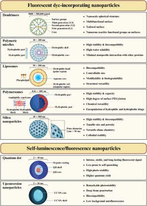
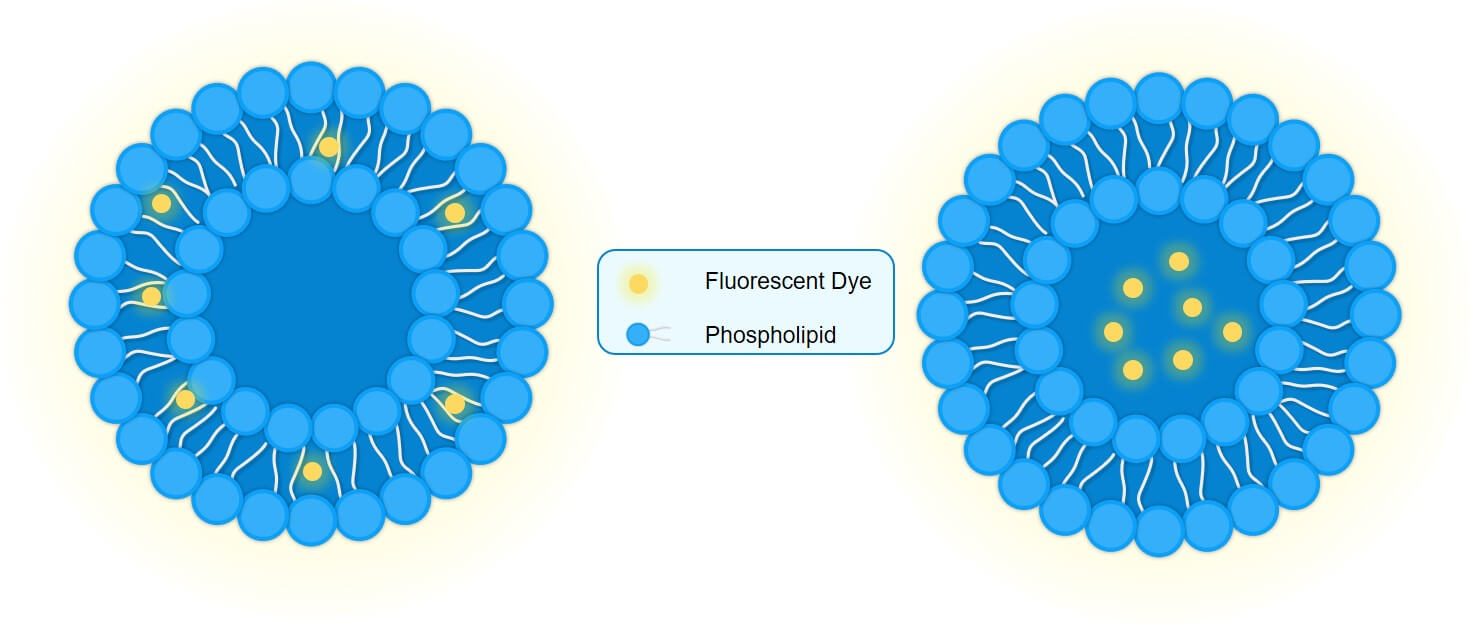
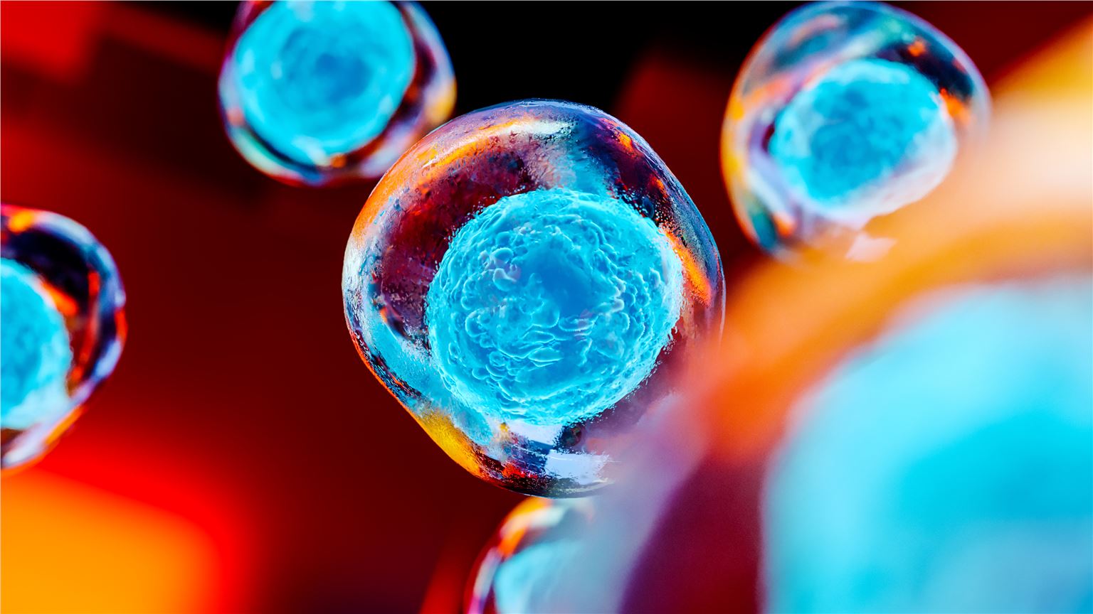
 Fig. 3 Various types of NPs in fluorescence imaging and their categorization into fluorescent dye-incorporating NPs and self-luminescence/fluorescence NPs.3,4
- Endosomal Escape: Visualize the release of payload from endosomes into the cytoplasm.
- Mechanism of Entry: Distinguish between clathrin-mediated endocytosis, caveolae-mediated uptake, or membrane fusion.
- Flow Cytometry: Quantify uptake efficiency across different cell populations with high-throughput precision.
Fluorescent Liposome Development Service for Targeted Drug Delivery
In the complex landscape of drug delivery, the ability to visualize the journey of a nanocarrier is as critical as the therapeutic payload itself. Tracking cellular uptake, intracellular trafficking, and systemic biodistribution requires high-fidelity imaging tools that do not compromise the integrity of the delivery system. At Creative Biolabs, we provide researchers with precision-engineered fluorescent liposomes that act as reliable beacons for your data.
Start Your Fluorescent Liposome Project
Background
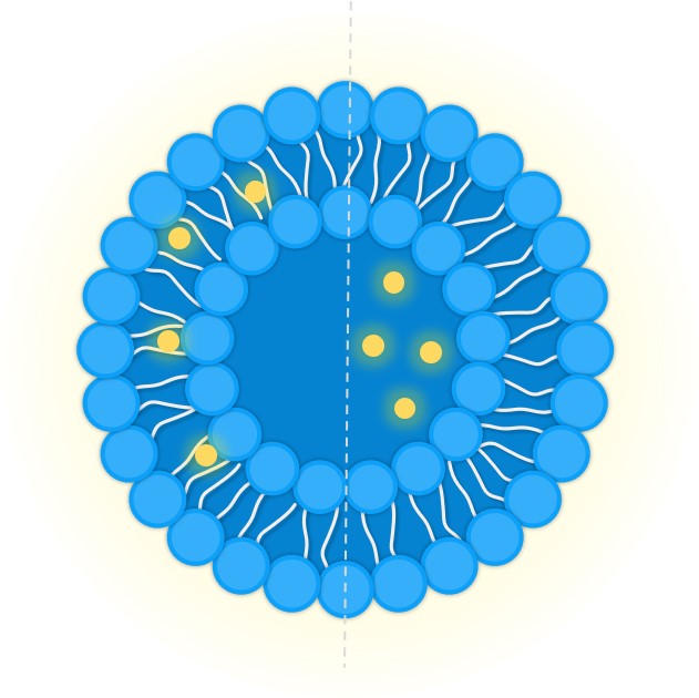
What Are Fluorescent Liposomes?
Fluorescent liposomes are phospholipid vesicles that have been modified with fluorescent probes either within the lipid bilayer or encapsulated inside the aqueous core. They serve as powerful tools for investigating the behavior of liposomal drug delivery systems in vitro and in vivo. By mimicking the physicochemical properties of therapeutic liposomes (size, charge, and surface chemistry), they provide a visual proxy for tracking drug delivery without the need for radioactive tracers.
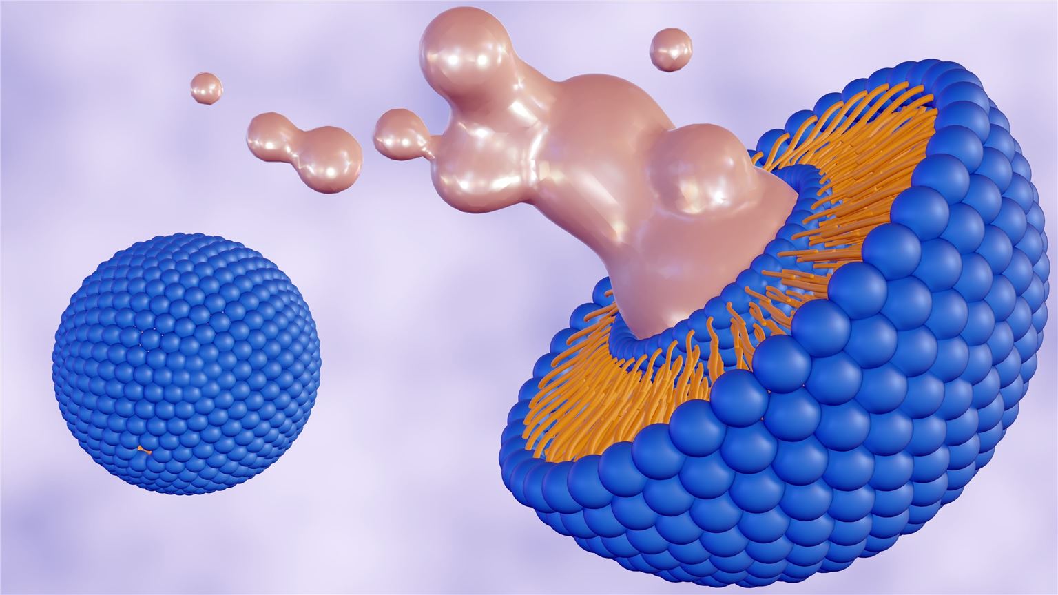
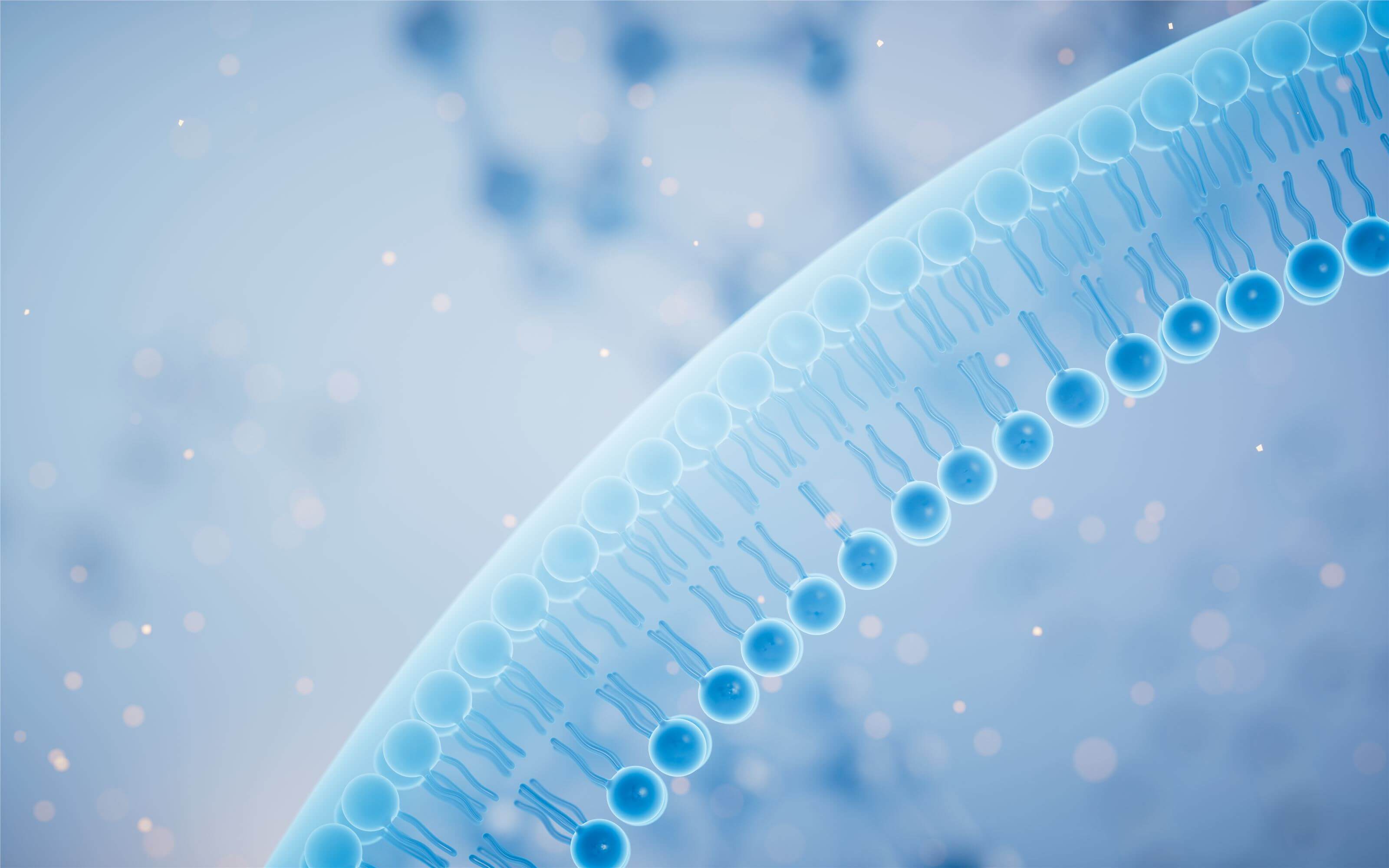
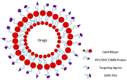 Fig. 1 Functionalized liposome for image-guided drug delivery.1,4
Why Fluorescent Liposomes Matter in Research
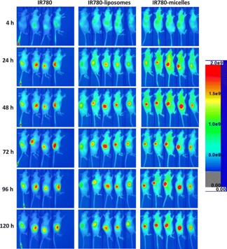 Fig. 2 In vivo NIRF imaging of fluorescent liposomes with Tumor model.2,4
Validating a targeted delivery system requires proof that the carrier actually reaches the intended site. Fluorescent liposomes allow researchers to:
- Confirm Targeting: Verify that ligand-conjugated liposomes bind specifically to target cells.
- Monitor Trafficking: Observe endosomal escape and subcellular localization.
- Assess Stability: Determine if the carrier remains intact in biological fluids prior to reaching the target.
Labeling Strategies
The placement of the fluorophore dictates the type of data obtained:
- Membrane Labeling: Lipophilic dyes (e.g., DiI, DiR) are intercalated into the lipid bilayer. This is stable and ideal for tracking the liposome carrier itself during cellular uptake and biodistribution studies.
- Aqueous Core Labeling: Hydrophilic dyes (e.g., Calcein, Carboxyfluorescein) are encapsulated in the interior. This strategy is essential for studying payload release, membrane fusion, and leakage kinetics (e.g., self-quenching assays).
Our Fluorescent Liposome Development Solutions
Our fluorescent liposome is designed to match your specific research goals, whether you are tracking endosomal escape or analyzing tissue accumulation.
Foundational Fluorescent Engineering
- Formulation Development & Optimization: We design robust lipid backbones (using DOTAP, DOPC, DSPC, Cholesterol, etc.) tailored to your stability requirements.
- Precise Dye Selection & Labeling: Whether you need membrane intercalation (using lipophilic dyes like DiI/DiD) or aqueous core encapsulation (using hydrophilic dyes like Calcein/FITC), we select the optimal fluorophore to match your imaging equipment.
- Physicochemical Adjustment: Fine-tuning of size, Zeta potential, and membrane fluidity to ensure the fluorescent control perfectly mimics your therapeutic candidate.
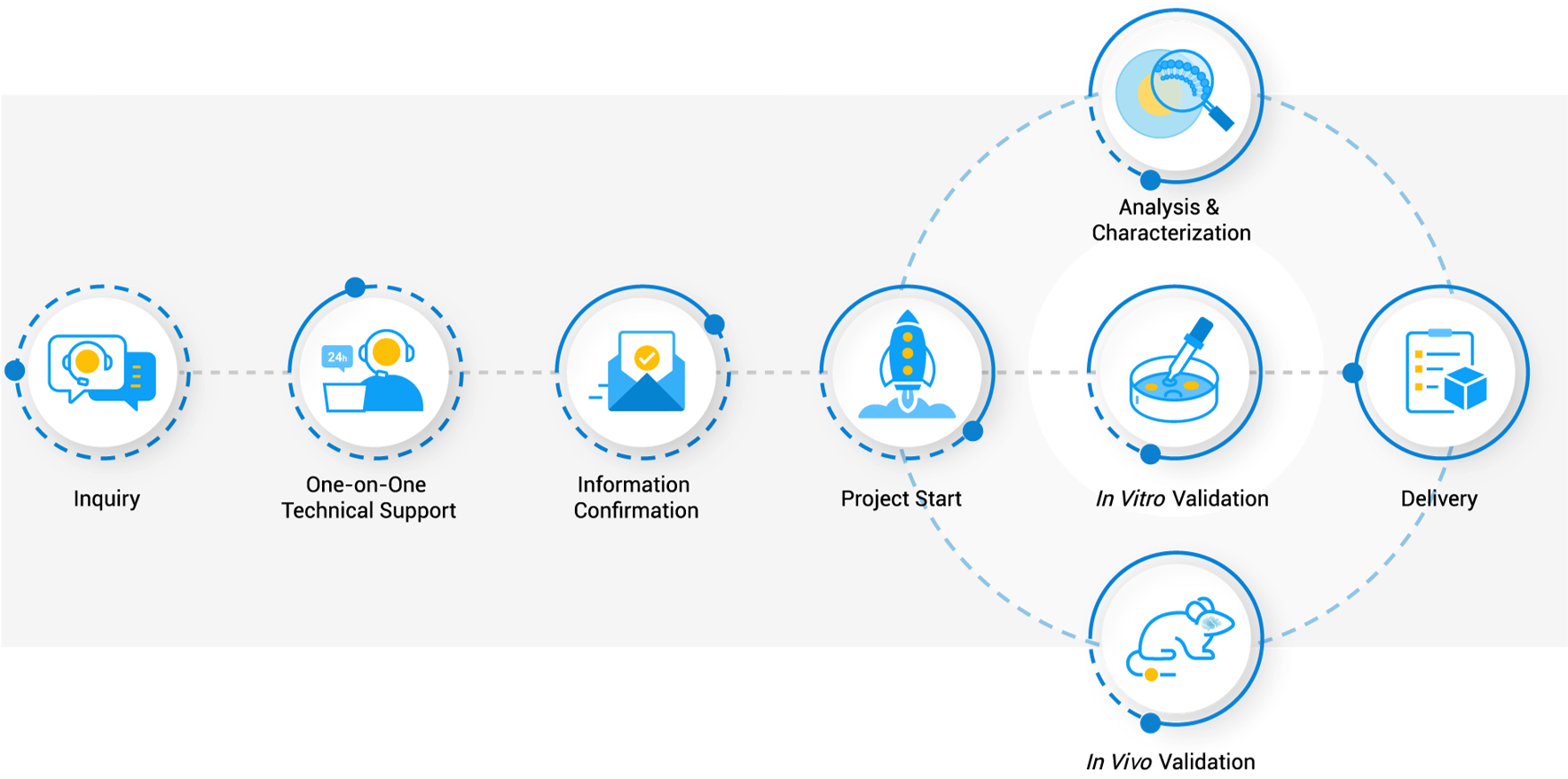
Workflow
Translating Nanocarrier Versatility into Therapeutic Efficacy
Our fluorescent liposomes are engineered to support a diverse array of experimental endpoints for Small Molecules, Nucleic Acids (siRNA, mRNA, CRISPR), and Proteins.
In Vitro Cellular Uptake & Trafficking
In Vivo Biodistribution & Imaging
- Tissue Accumulation: Track the accumulation of liposomes in target organs (e.g., liver, tumor microenvironment) vs. off-target tissues.
- Near-Infrared (NIR) Imaging: Utilizing dyes like DiR or Cyanine 7 (Cy7) for deep-tissue imaging with minimal autofluorescence background.
- Theranostics: Co-encapsulate therapeutic agents with imaging probes to monitor drug delivery and therapeutic response simultaneously
Why Choose Creative Biolabs?
High-Fidelity Signal
We utilize high-purity fluorophores and optimized loading techniques to prevent rapid photobleaching and self-quenching issues.
Biologically Relevant Controls
Our formulations are designed to mimic the physical behavior of drug-loaded liposomes, ensuring your control data is valid.
Custom Conjugation Expertise
We possess deep experience in complex surface chemistry, allowing for the precise attachment of sensitive biological ligands without loss of activity.
Scalability & Consistency
From small-scale R&D batches to larger pilot lots, we ensure strict batch-to-batch consistency in size (PDI < 0.2) and fluorescence intensity.
Turnkey Solutions
We provide ready-to-use reagents that save your lab time, allowing you to focus on biology rather than formulation chemistry.
Creative Biolabs stands at the forefront of nanomedicine development, offering robust solutions that illuminate the complex interactions between drug carriers and biological systems. Ready to visualize your success? Our team of formulation scientists is ready to discuss your specific imaging needs and design a liposome strategy that fits your project goals.
Related Services & Products
Related Services
Related Products
| Product Name | Description | Inquiry |
|---|---|---|
| Functionalized Fluorescent Dyes | High-purity lipid-dye conjugates such as DSPE-PEG-FITC, DSPE-PEG-Rhodamine, and DSPE-PEG-Cy5 for self-assembly. | |
| Membrane Labeling Reagents | Specialized lipids labeled with lipophilic tracers like DiD, DiI, and DiR for stable membrane integration. | |
| Encapsulated Controls | Pre-formed liposomes with high-concentration aqueous dyes (FITC, Calcein) for leakage and fusion assays. | |
| Theranostic Liposomes | Drug-loaded fluorescent liposomes capable of co-delivering therapeutics (Doxorubicin, Paclitaxel) and imaging agents. |
FAQs
Can I request a specific fluorophore that matches my microscope's filter set?
Yes, we can incorporate almost any commercially available hydrophobic or hydrophilic fluorophore. Common requests include FITC, Rhodamine B, DiI, DiD, DiR, and Cy5/Cy7.
What is the stability of your fluorescent liposomes?
Liquid formulations are typically stable for 3-6 months at 4°C. Our lyophilized formulations can be stored at -20°C for up to 12 months with no loss of integrity upon reconstitution.
Can you make fluorescent liposomes that mimic my specific drug formulation?
Absolutely. We can match the lipid composition, size, and surface charge (Zeta potential) of your specific therapeutic candidate to ensure the biodistribution data is translatable.
Do you offer sterile formulations for cell culture?
Yes, all our formulations intended for biological use are passed through a 0.22 μm filter and prepared under aseptic conditions.
What is the typical size range you can achieve?
We can precisely engineer liposomes from 50 nm up to 200 nm for systemic delivery, and larger micron-sized vesicles (GUVs) for specific mechanism studies.
References
- Lamichhane N, Udayakumar T S, D'Souza W D, et al. Liposomes: clinical applications and potential for image-guided drug delivery[J]. Molecules, 2018, 23(2): 288. https://doi.org/10.3390/molecules23020288
- Li, Shihong, et al. "Near infrared fluorescent imaging of brain tumor with IR780 dye incorporated phospholipid nanoparticles." Journal of translational medicine 15.1 (2017): 18. https://doi.org/10.1186/s12967-016-1115-2.
- Han, Chae Yeon, et al. "Nano-fluorescence imaging: advancing lymphatic disease diagnosis and monitoring." Nano Convergence 11.1 (2024): 53. https://doi.org/10.1186/s40580-024-00462-1.
- Distributed under Open Access license CC BY 4.0, without modification.
Our services are For Research Use Only. We do not provide services to individuals.
Online Inquiry