We collaborate to define your project goals, selecting the most relevant cell models (e.g., HUVEC, microvascular ECs) and optimizing the assay format.
Endothelial Cell Migration & Invasion Analysis Service
Are you currently facing long drug development cycles, inconsistent data from manual scratch assays, or difficulty in quantifying the invasive potential of your anti-angiogenic candidates? Endothelial cell migration & invasion analysis service at Creative Biolabs helps you obtain high-quality kinetic data and develop highly specific therapeutic antibodies through real-time impedance-based monitoring, automated 3D matrix invasion platforms, and expert-led physiological modeling. We solve the challenge of complex motility quantification.
Overview What We Can Offer Workflow Required Materials Highlights Publication Customer Reviews FAQs Related Services
Overview
Endothelial cell motility is a fundamental requirement for angiogenesis, tissue regeneration, and inflammatory responses. Migration involves the collective or individual movement of cells across surfaces, while invasion signifies the active degradation of extracellular matrix (ECM) barriers. Creative Biolabs leverages these insights to provide highly credible, validated assay platforms.
We utilize a multi-pronged strategic approach to capture the full spectrum of endothelial motility. Our methodology distinguishes between chemotaxis (movement toward soluble gradients) and haptotaxis (movement toward matrix-bound cues). For basic screening, we employ high-density monolayer wound healing models to assess collective cell movement and front-rear polarization. For advanced oncology and regenerative research, we utilize 3D invasion strategies that incorporate standardized basement membrane extracts. This allows us to observe the proteolytic activity of "tip cells" as they navigate complex fiber densities, ensuring the data we generate is representative of the in vivo environment.
Empower Your Research with Creative Biolabs — Begin Your Expert Consultation Today
What We Can Offer
Real-Time Kinetic Monitoring
Continuous, label-free assessment of cell migration using impedance-based technology for minute-by-minute data acquisition.
3D Matrix Invasion Assays
Specialized Transwell-based systems coated with Matrigel or collagen to evaluate the proteolytic and invasive capacity of cells.
Automated Scratch Assays
Highly reproducible wound-healing models created with standardized mechanical or optical tools to measure collective migration rates.
Chemotactic Gradient Modeling
Precision setup of growth factor gradients (e.g., VEGF, bFGF) to study directional cell translocation and filopodia dynamics.
Multi-Parameter Image Analysis
Quantification of migration velocity, persistence, and directionality using advanced single-cell tracking software.
Pharmacological Profiling
Determination of IC50/EC50 values for novel inhibitors or stimulators of the invasive cascade.
Workflow
Study Design and Cell Selection
Substrate and Matrix Preparation
Standardized ECM components are applied to culture surfaces or Transwell inserts, ensuring a uniform barrier for invasion or a consistent substrate for migration.
Cell Synchronization and Seeding
Cells are synchronized via serum starvation and seeded at precise densities to ensure that movement, not proliferation, is the measured variable.
Compound Treatment and Gradient Setup
Test agents are introduced alongside positive and negative controls, while stable chemoattractant gradients are established to drive directional movement.
Automated Data Acquisition
Assays are placed in climate-controlled imaging or impedance systems, capturing data at frequent intervals to track the dynamic progression of cell motility.
Quantitative Reporting and Analysis
Our experts extract key parameters such as T-half, peak migration rate, and invasion ratios, providing a detailed final report with statistical validation.
Required Starting Materials
- Test Agent Profiles: Documentation regarding the solubility, stock concentration, and handling requirements of your compounds or biologics.
- Target Bio-Benchmarks: Information on specific growth factors or known inhibitors you wish to include as reference controls for the assay.
Highlights
Superior Label-Free Analytical Precision
Our advanced impedance-based monitoring systems eliminate the absolute need for exogenous fluorescent dyes or metabolic tracers, preventing any potential alteration of natural cell behavior and ensuring exceptionally high cellular viability throughout the duration of the assay.
Comprehensive Full Kinetic Curve Resolution
By capturing the entire temporal duration of the migratory process from start to finish, we identify transient drug effects, lag phases, and optimal efficacy windows that traditional single-point endpoint measurements frequently overlook in standard protocols.

Advanced AI-Driven Image Segmentation
We utilize proprietary deep-learning software to provide strictly objective, single-cell tracking and collective migration metrics, effectively reducing inter-operator variability and human bias to negligible levels for cleaner data sets.
Physiologically Relevant 3D Barrier Invasion
Our invasion models accurately simulate the complexity of the vascular basement membrane using tunable fiber density and matrix compositions, providing a rigorous and translatable test for anti-metastatic drug candidates.
Experience the Creative Biolabs Difference — Contact Us for a Tailored Quote
Publication
Endothelial cells regulate breast cancer invasion through metabolic interactions that influence tumor cell energy supply. When glucose availability was restricted, endothelial cell–dependent tumor invasion into the surrounding matrix was markedly reduced, indicating that adequate glucose metabolism is required for endothelial-driven migratory responses. In parallel, inhibition of ATP synthase impaired mitochondrial ATP production and further suppressed invasion toward vascular structures. These metabolic constraints weakened the supportive role of endothelial cells in guiding tumor cell movement, resulting in diminished directional invasion. By limiting both glycolytic input and oxidative phosphorylation, energy stress was imposed on the microenvironment, revealing a strong dependence of endothelial-mediated tumor invasion on active energy-generating pathways. This highlights metabolic regulation as a critical factor in endothelial–tumor cell interactions during invasive progression.
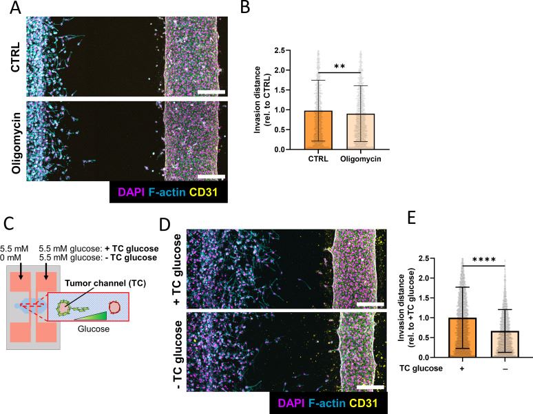 Fig.1 Glucose limitation and ATP synthase blockade suppress endothelial-driven tumor invasion.1
Customer Reviews
- "[Superior Kinetic Clarity] Using Creative Biolabs' endothelial cell migration & invasion analysis service in our research has significantly improved our understanding of how our peptide affects early-stage migration. The real-time tracking revealed an inhibitory effect within the first three hours that was invisible in our previous 24-hour end-point assays." -18 days ago, D***r M.
- "[High-Resolution Invasion Data] Using Creative Biolabs' Endothelial cell migration & invasion analysis service in our research has significantly facilitated our anti-angiogenesis antibody screening. The automated 3D invasion index provided the statistical power we needed to move our lead candidate into pre-clinical animal models." -1 month ago, S***n P.
- "[Reliable Collective Migration Modeling] Using Creative Biolabs' endothelial cell migration & invasion analysis service in our research has significantly facilitated our study of diabetic wound healing factors. The reproducibility of their automated scratch assay allowed us to compare multiple growth factor variants with total confidence." -5 days ago, A***a L.
FAQs
How do you ensure that you are measuring migration and not cell proliferation?
We utilize multiple strategies, including serum starvation and the use of mitotic inhibitors like Mitomycin C. Furthermore, our real-time analysis detects movement within hours, a timeframe significantly shorter than a typical cell division cycle.
Can your invasion assay simulate the blood vessel wall for transmigration studies?
Yes, we can establish a confluent endothelial monolayer on the Transwell membrane to study trans-endothelial migration (TEM), a process crucial for understanding cancer metastasis and inflammatory responses.
What is the advantage of impedance monitoring over traditional staining?
Impedance monitoring is label-free and provides continuous data acquisition. Traditional staining is an end-point assay that requires fixing the cells, which loses vital kinetic data and can introduce processing artifacts.
Can I provide my own specific extracellular matrix (ECM) for the invasion assay?
While we offer standardized matrices like Matrigel and Collagen, we can optimize protocols to include customer-provided matrices or custom hydrogels to meet highly specific research needs.
Related Services
3D Tumor Vascular Network Bioprinting & Perfusion Evaluation Service
This service establishes three-dimensional vascularized tumor models to assess network formation, perfusion capacity, and functional response within complex tissue-like environments.
Learn More →In Vivo Vascular Network Formation & Matrigel Plug Assay Service
This service evaluates angiogenic responses in vivo through established implantation-based models, supporting confirmation of vascular growth and functional integration under physiological conditions.
Learn More →Our scientific team is ready to help you design a tailored study for your specific vascular motility research. Whether you need a high-throughput migration screen or a complex 3D invasion model, Creative Biolabs offers the support and expertise your project demands.
Contact Our Team for More Information on Endothelial Cell Migration & Invasion Analysis Service and to Discuss Your Project.
Reference
- Tan, Matthew L et al. "Endothelial cells metabolically regulate breast cancer invasion toward a microvessel." APL bioengineering vol. 7,4 046116. 4 Dec. 2023. Distributed under Open Access license CC BY 4.0, without modification. https://doi.org/10.1063/5.0171109
 Download our brochure
Loading case studies...
Online Inquiry