Fluorescence Lifetime Imaging Microscopy (FLIM) Service for Organelle Metabolic Research
A New Method for Metabolic Cofactor Imaging
The intrinsic fluorescence of the metabolic cofactors NADH and NADPH has been used as an indicator of the metabolic state of living tissues for more than 50 years. With the help of modern developments in laser-scanning microscopy, pulsed laser sources, and time-resolved fluorescence detection, Creative Biolabs is pleased to measure the fluorescence lifetime of metabolic cofactors within cells and tissues using fluorescence lifetime imaging microscopy (FLIM). Our strategy is highly sensitive and will provide information on the metabolic state of complex biological preparations at a molecular level. Free and protein-bound forms of metabolic cofactors will be imaged and the redox state in live cells will be calculated efficiently.
The metabolism of mitochondria contributes significantly to the survival and proliferation of cancer cells, which also coexists with increased glycolytic activity. With years of experience in organelle metabolic research, we also offer mitochondrial activity measurement services to characterize cancer metabolism patterns, identify metabolic vulnerabilities, and identify new drug targets.
Analysis of Tumor Heterogeneity by FLIM
Moreover, to assess progression and tailor treatment for a tumor, it is of vital importance that tumor heterogeneity be detected and quantified. In recent years, considerable progress has been achieved in understanding the existence of a high degree of spatial metabolic heterogeneity within tumors due to molecular characterization and metabolic profiling. In addition to basic aspects of carcinogenesis, there are also important clinical applications for mapping and quantification of metabolic heterogeneity at the cell level.
FLIM of metabolic cofactors allows one to probe cellular metabolism by measuring their fluorescence lifetime in live cells and tissues non-invasively, and the contrast between the different structures also can be observed. Whether there is a relationship between cancer heterogeneity, cancer progression, and metabolic cofactors signal intensity is paid more and more attention. It is needed to validate these autofluorescent metabolites as cancer metabolic biomarkers. Based on our label-free FLIM with image acquisition and (sub)cellular resolution, we will gain insight into the metabolic heterogeneity of cancer cells. Creative Biolabs is proud to launch our FLIM-based tumor heterogeneity analysis service to better identify molecules and their interaction with other molecules.
Advantages of Our FLIM Service
- Imaging free and protein-bound metabolic cofactors
- Extremely sensitive
- (Sub)cellular resolution
- Analysis of metabolic heterogeneity of cancer cells
- Observation of the contrast between the different structures
Publications Sharing
Publication 1
Technology Used: FLIM with two-photon microscopy
Journal: PLoS Pathogens
IF: 6.7
Research Findings: The authors visualized the metabolic pathogen-host interactions during intracellular Chlamydia trachomatis infections with high spatial and temporal resolution in living cells by FLIM with two-photon microscopy. The findings show that intracellular chlamydial metabolism has a direct association with the cellular NADH signaling pathways, which have been associated with host cell survival and longevity.
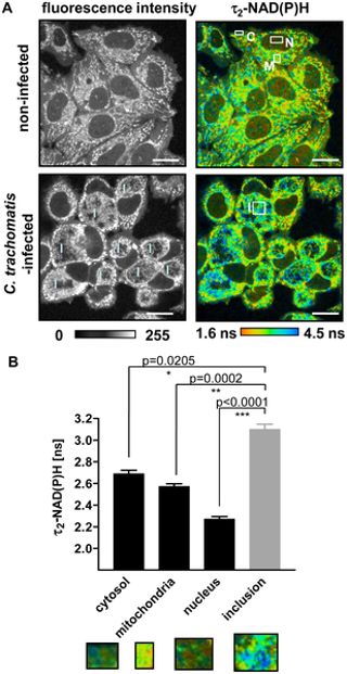 Fig.1 FLIM of NAD(P)H by two-photon microscopy.1
Except for Tumor Metabolism Characterization, we also provide:
- Functional Genetic Screening for Tumor Metabolism
- Tumor Model Construction for Cancer Metabolism Research
- In Vivo Analysis for Tumor Metabolism
If you need our help in Tumor Energy Metabolism Analysis, please feel free to contact us.
Reference
- Szaszák, Márta, et al. "Fluorescence lifetime imaging unravels C. trachomatis metabolism and its crosstalk with the host cell." PLoS Pathogens 7.7 (2011): e1002108. Distributed under Open Access license CC BY 4.0, without modification.
 Download our brochure