pH Detection Service under Tumor Metabolic Microenvironment
pH in Tumor Metabolic Microenvironment
Changes in the metabolism of tumor cells cause the pH of the tumor microenvironment (TME) to become acidic. This alteration is characteristic of abnormal cell-cell interactions and disruption of homeostasis. In this state, tumor cells preferentially utilize glycolysis rather than oxidative phosphorylation as the primary means of energy release. This change results in an approximately tenfold increase in lactate load in the extracellular environment, with diffusion transport of H+ ions to the interstitium.
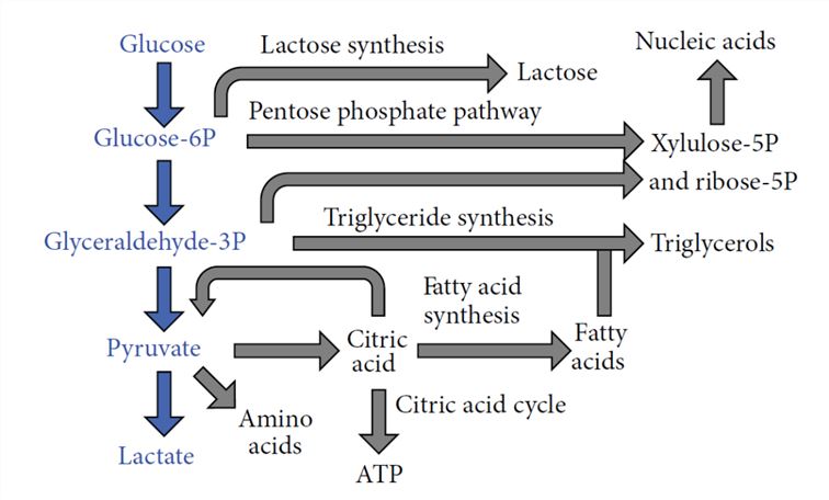 Fig.1 Glycolysis and related metabolic pathways (production of tumor growth molecules).1
- pH is a commonly used parameter in TME studies. The success of targeting the pH of the tumor microenvironment depends in part on the accurate detection of the pH of the tumor microenvironment, and the methods now used include PET radiotracer, MR spectroscopy, MRI, and other techniques.
- Relying on advanced instrumentation and professional staffing, Creative Biolabs provides a series of detection methods that target the pH of the tumor microenvironment, providing a strong basis for your tumor metabolism research.
Our Service
- Imaging for pH Detection
Bioimaging is a very effective technique to study the tissue structure and physiological function of organisms, with the advantages of high sensitivity, non-invasive detection, high selectivity, and real-time imaging, so bioimaging is a powerful tool for monitoring important molecules in organisms. Creative Biolabs offers, but is not limited to, the following imaging approaches:
- Table 1 Imaging for pH Detection.1
| Methods | Features |
|---|---|
| Microelectrode |
|
| Fluorescence imaging |
|
| PET |
|
| MRS |
|
| pH dependent T1 relaxation |
|
| CEST MRI |
|
The following is published data for pH detection using image methods:
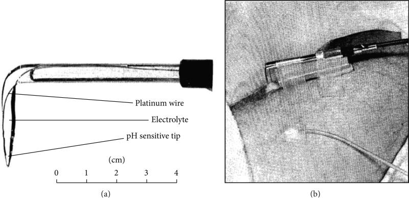 Fig.2 pH microelectrode imaging.1
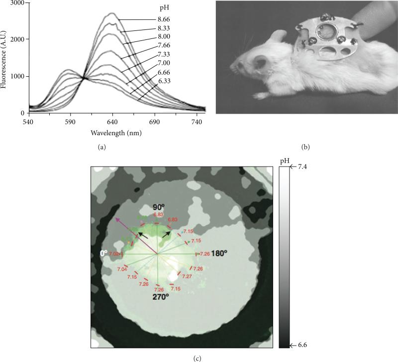 Fig.3 Fluorescence imaging.1
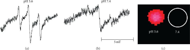 Fig.4 EPR imaging.1
- Electrochemistry for pH Detection
This is a novel design that utilizes the conductive polymer polyaniline as a sensor in combination with an optimized electrode. This electrochemical method allows real-time detection of pH changes outside cancer cells.
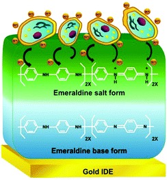 Fig.5 Schematic diagram of how the sensor works.2
Features of this method:
- Real-time detection of extracellular pH
- High-sensitivity diagnosis of cancer cells
- Real-time evaluation of glycolysis inhibition efficiency of anticancer drugs
In addition to pH testing, Creative Biolabs provides a series of Tumor Metabolic Microenvironment Analysis services through continuous in-depth research on tumor metabolism. If you are interested, please do not hesitate to contact us.
References
- Chen, L.Q. and Mark D.P. "Evaluating pH in the extracellular tumor microenvironment using CEST MRI and other imaging methods." Advances in radiology. (2015): 206405.
- Thakur, B.; et al. "Probing extracellular acidity of live cells in real time for cancer detection and monitoring anti-cancer drug activity." Chemical communications. (2015): 7015-8.
 Download our brochure