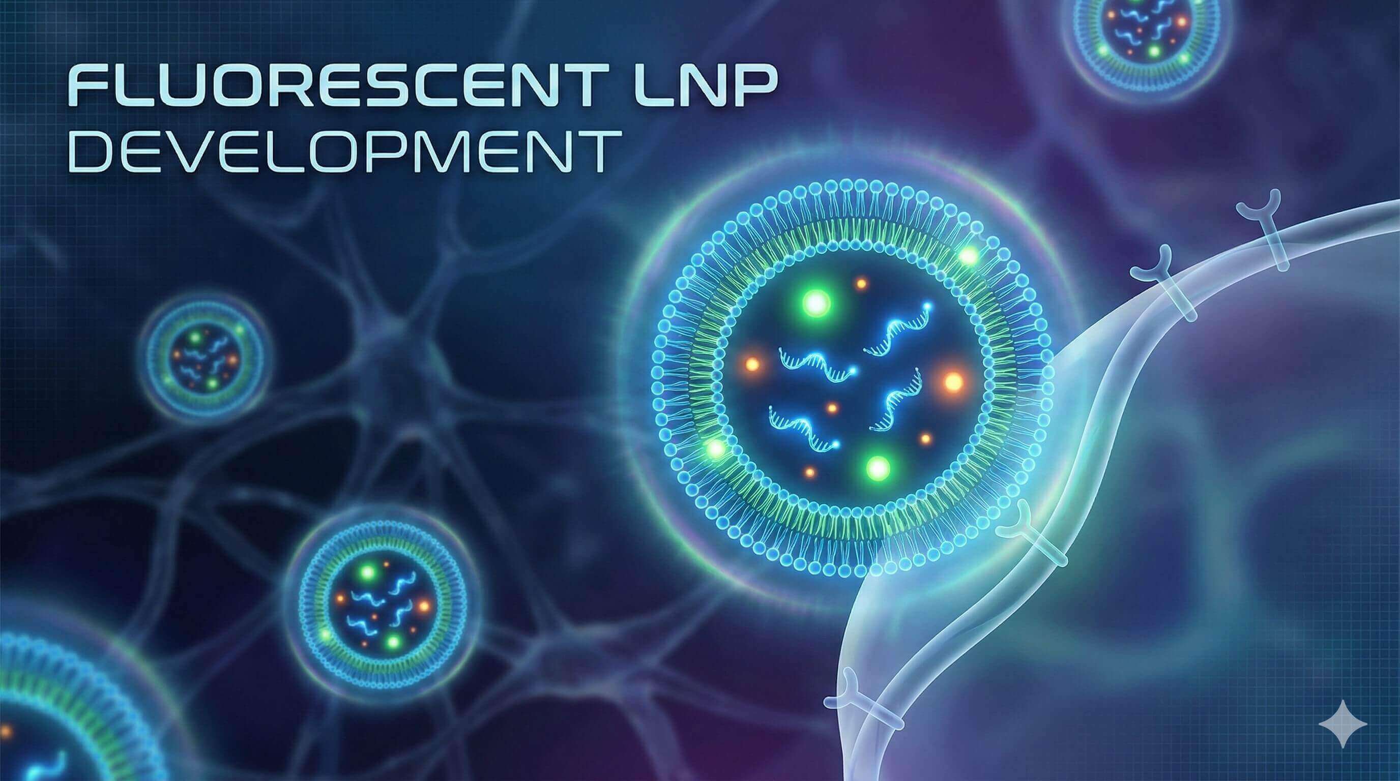
Fluorescent LNP Development Service for Targeted Delivery
Understanding the precise biological fate of Lipid Nanoparticles (LNPs)—from systemic circulation to cytosolic release—often remains a "black box" in preclinical development. Creative Biolabs' fluorescent LNP development service transforms this uncertainty into actionable data. By utilizing fluorescent mRNA or fluorescent labeling of the LNP, we empower researchers to visualize cellular uptake, track intracellular trafficking, and quantify biodistribution with exceptional sensitivity.
Start Your Custom Fluorescent LNP Project
The Science of Visualization: Unlocking LNP Behavior
The Crucial Role of Visualization in Nanomedicine
Lipid Nanoparticles (LNPs) are complex systems whose behavior in biological environments is dynamic and often unpredictable. While physicochemical properties (size, PDI) can be measured in vitro, understanding their true biological fate requires direct observation. Fluorescent LNPs serve as indispensable tools to bridge the gap between formulation chemistry and biological function, providing critical insights into:
Biodistribution Profiling: Visualizing systemic accumulation in filter organs (liver, spleen) versus target tissues (e.g., tumors, brain) to validate tissue specificity and off-target effects.
This visual data is essential for distinguishing between delivery failure and therapeutic inefficacy, enabling rational design optimization.
Precision Engineering Approaches for LNP Visualization
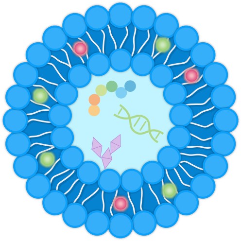
Achieving high-resolution biological data requires a labeling strategy that is rigorously aligned with the specific research objective. At Creative Biolabs, we have developed a bifurcated engineering framework to address this need, offering distinct yet complementary methodologies: Fluorescent mRNA for payload tracking and Fluorescent Labeling of LNP for vehicle characterization. This targeted approach ensures optimal signal fidelity and mechanistic clarity.
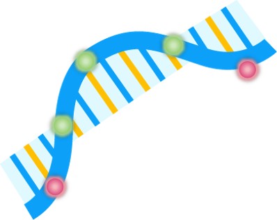
This strategy focuses on the payload itself. By using chemically modified nucleotides or reporter genes, we can track the genetic material independently of the lipid carrier.
Mechanism: Fluorophores (e.g., Cy5, Cy3) are covalently attached to the mRNA backbone, or the mRNA encodes a fluorescent reporter protein (e.g., EGFP).
Application: Essential for studying endosomal escape, cargo release, and protein expression efficiency.
Advanced Visualization Technologies & Analytical Readouts
To translate fluorescent signals into actionable biological insights, Creative Biolabs utilizes a comprehensive suite of high-resolution imaging and analytical platforms. These technologies allow for the rigorous quantification of LNP performance from the cell level to whole-organism biodistribution.
| Visualization Technique | Key Application | Analytical Capabilities & Readouts |
|---|---|---|
| Multiparametric Flow Cytometry (FACS) | Quantitative Uptake Analysis | High-throughput quantification of Mean Fluorescence Intensity (MFI) and % Positive Cells. Enables precise determination of cellular uptake kinetics and transfection efficiency across heterogeneous cell populations. |
| Confocal Laser Scanning Microscopy (CLSM) | Subcellular Trafficking & Colocalization | High-resolution Z-stack imaging to visualize intracellular fate. Critical for calculating Colocalization Coefficients to distinguish between endosomal entrapment and cytosolic release (endosomal escape). |
| In Vivo Imaging Systems (IVIS) | Whole-Body Biodistribution | Non-invasive, longitudinal tracking of NIR-labeled LNPs. Provides quantitative data on Total Radiant Efficiency and Tissue-to-Background Ratios, mapping accumulation in filter organs (liver, spleen) versus target tissues. |
| High-Content Screening (HCS) | Automated Phenotypic Profiling | Automated image-based analysis to correlate LNP uptake with cellular phenotypic changes (e.g., cytotoxicity, morphological shifts) at scale, enabling high-throughput formulation ranking. |
Precision Engineering for Bioimaging
Creative Biolabs offers a comprehensive suite of fluorescent formulation services, ranging from rapid screening batches to scaled-up, sterile preparations for animal studies. We provide a "turnkey" solution that integrates our Module Delivery Systems with high-quantum-yield fluorophores.
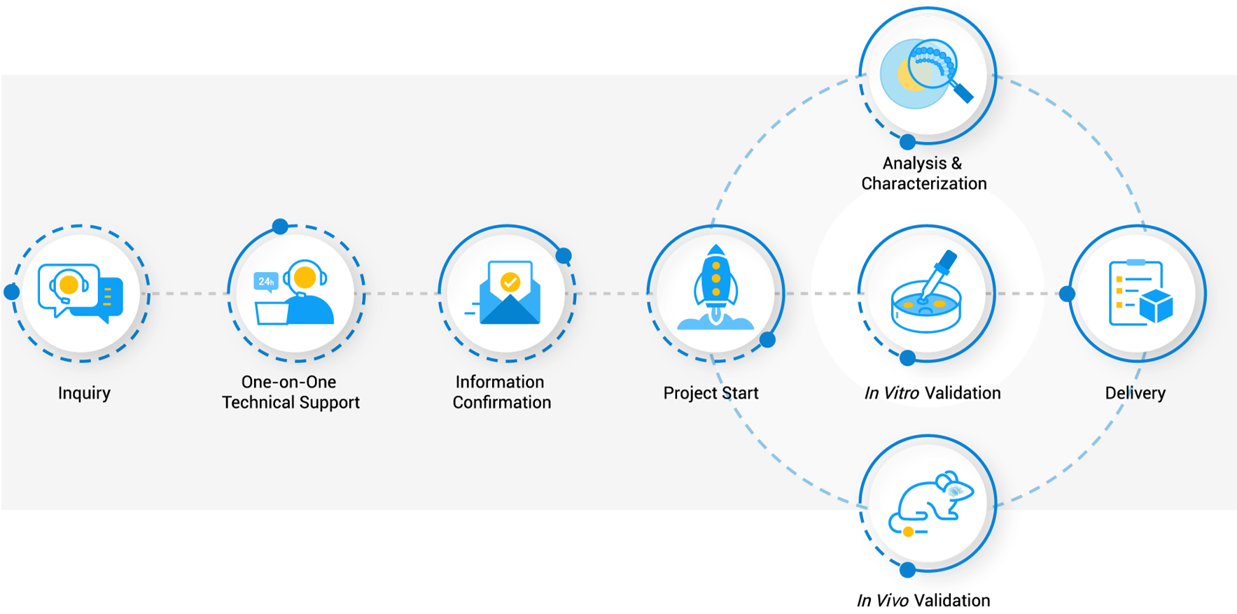
Workflow
Applications: Illuminating Biological Pathways
Validating Intracellular Trafficking and Endosomal Escape
The primary barrier to RNA delivery is entrapment within endosomes. By utilizing pH-sensitive probes or dual-labeling strategies, researchers can visually distinguish between LNPs trapped in lysosomes and functional cargo released into the cytosol, providing definitive proof of mechanism.
In Vivo Biodistribution and Tissue Targeting
Using Near-Infrared (NIR) dyes like DiR or Cy7, our LNPs are optimized for whole-animal imaging (e.g., IVIS). This allows for non-invasive longitudinal monitoring of LNP accumulation in deep tissues, crucial for evaluating the efficacy of tissue-specific targeting modules (e.g., targeting hepatocytes vs. splenocytes).
Flow Cytometry-Based Uptake Quantification
For high-throughput screening, our fluorescent LNPs enable the precise quantification of transfection efficiency across different cell populations. This is essential for comparing the potency of different Targeted Modules (aptamers vs. peptides) in mixed cell cultures.
Why Choose Creative Biolabs?
Preserved Physicochemical Integrity
We optimize dye-to-lipid ratios to ensure that fluorescence labeling does not destabilize the particle or significantly alter the size (~100 nm) and PDI (<0.2).
High-Efficiency Encapsulation
Our microfluidic platform maintains high encapsulation efficiency (>85%) even with the addition of hydrophobic dyes.
Broad Fluorophore Library
We offer a wide range of excitation/emission options (Green, Red, Far-Red, NIR) compatible with standard confocal microscopes and in vivo imaging systems.
Unlike standard vendors, we seamlessly integrate active targeting ligands (antibodies, peptides) onto fluorescent particles, allowing you to validate your targeting hypothesis directly.
Stability Solutions
Our optional lyophilization service ensures your fluorescent reagents remain stable for shipping and storage without dye leakage or aggregation.
Creative Biolabs provides the clarity your research demands. By combining precision microfluidics with advanced fluorescent technologies, we deliver LNP formulations that are not only therapeutic vehicles but powerful analytical tools. Whether you are validating a new targeting ligand or profiling the biodistribution of a novel mRNA vaccine, our Fluorescent LNP Development Service provides the visual evidence required to advance your pipeline with confidence.
Related Services & Products
Related Services
Chemical Synthesis Services
Nucleic Acid Synthesis Services
Related Products
| Product Name | Description | Inquiry |
|---|---|---|
| EGFP mRNA-LNP | Ready-to-use LNPs encapsulating EGFP mRNA, serving as a positive control for transfection and expression studies. | |
| Fluc mRNA-LNP | LNPs encapsulating Firefly Luciferase mRNA. Perfect for sensitive bioluminescence imaging of in vivo expression. | |
| Custom Fluorescently Labeled LNP | Build-your-own LNP service. Select payload (Empty, siRNA, mRNA) and Fluorophore (DiI, DiO, DiR, Cy5, etc.). | |
| RNA-LNP Encapsulation Assay Kit | A complete kit using RiboGreen reagents for the precise quantification of Encapsulation Efficiency (EE%). | |
| Lipophilic Tracer Dyes | High-purity DiI, DiO, DiD, and DiR dyes optimized for membrane incorporation in liposomes and LNPs. |
FAQs
Will adding a fluorescent dye change the size or charge of my LNP?
At Creative Biolabs, we carefully optimize the dye-to-lipid ratio to ensure that the incorporation of the fluorophore has a negligible impact on the particle's hydrodynamic diameter, PDI, and Zeta potential, preserving the formulation's native biological behavior.
Can I label both the lipid shell and the RNA cargo simultaneously?
Yes. We specialize in Dual-Labeled LNPs. We can incorporate a lipophilic dye (e.g., DiI) into the lipid shell and encapsulate a fluorescent mRNA (e.g., EGFP mRNA). This is highly recommended for studying cargo release kinetics and mechanism of action.
Which dye is best for in vivo animal imaging?
For in vivo biodistribution studies, we recommend Near-Infrared (NIR) dyes such as DiR or Cy7. These fluorophores emit at wavelengths (>700 nm) that penetrate tissue more effectively and have lower autofluorescence background compared to green or red dyes.
Can you conjugate targeting ligands to fluorescent LNPs?
Absolutely. We can couple antibodies, peptides, or aptamers to the surface of fluorescent LNPs. This allows you to visually validate the specificity of your targeting strategy by comparing uptake in receptor-positive vs. receptor-negative cells.