Based on its advanced chemokine production measurement service, Creative Biolabs provides a comprehensive platform for chemokine production assays. Their skilled scientists use powerful technology to meet diverse customer needs and are ready to assist with any project.
Learn More →Identification, Quantification & Comparation Service of Leukocyte Population
Background Markers Technologies Publication Why Choose Us FAQs Customer Review Related Services Contact Us
Precision Insights into Leukocyte Dynamics
Creative Biolabs offers comprehensive services to analyze and characterize leukocyte populations and subpopulations, including lymphocytes, monocytes, and granulocytes, using advanced technologies. Our technologies include:
Flow Cytometry
Immunohistochemistry (IHC)
Markers of Leukocyte Populations
| Lymphocytes | Marker | Description |
|---|---|---|
| T Cells | CD3 | Pan-T cell marker, present on all T lymphocytes. |
| CD4 | Expressed on Helper T cells, crucial for antigen presentation. | |
| CD8 | Found on Cytotoxic T cells, involved in killing infected cells. | |
| CD25 | Marker of activated T cells, also expressed on regulatory T cells. | |
| CD45RO | Marker for memory T cells, distinguishing them from naive T cells. | |
| B Cells | CD19 | Pan-B cell marker, present on all B lymphocytes. |
| CD20 | Commonly used marker for B cells, also a target in some B cell malignancies. | |
| CD21 | Complement receptor, found on mature B cells. | |
| CD27 | Marker for memory B cells, indicating past antigen exposure. | |
| NK Cells | CD56 | Principal marker for Natural Killer (NK) cells. |
| CD16 | Low-affinity Fc receptor, involved in antibody-dependent cellular cytotoxicity (ADCC). | |
| Cell Type | Marker | Description |
| Monocytes | CD14 | Classical monocyte marker, binds to lipopolysaccharides (LPS). |
| CD16 | Found on non-classical and intermediate monocytes, associated with inflammation. | |
| CD64 | High-affinity Fcγ receptor, expressed on monocytes and macrophages. | |
| HLA-DR | Major histocompatibility complex class II molecule, indicating monocyte activation. | |
| Granulocytes | Marker | Description |
| Neutrophils | CD15 | Marker for granulocytes, prominently expressed on neutrophils. |
| CD66b | Specific to neutrophils, involved in adhesion and activation. | |
| CD16 | Low expression on neutrophils, involved in ADCC (higher in some conditions). | |
| Eosinophils | CD193 (CCR3) | Chemokine receptor specific to eosinophils, involved in recruitment to tissues. |
| CD125 | Interleukin-5 receptor alpha chain, expressed on eosinophils. | |
| CD9 | Integrin and adhesion molecules found on eosinophils. | |
| Basophils | CD123 | Interleukin-3 receptor alpha chain, a distinguishing marker for basophils. |
| CD203c | Activation marker on basophils, increases during allergic reactions. | |
| CD49b | Integrin alpha 1, found on basophils, involved in cell adhesion. |
Technologies
- Flow Cytometry Analysis
Our flow cytometry services provide detailed analysis of leukocyte populations based on surface markers and functional characteristics. This technique allows for high-throughput, multiparameter analysis of immune cells.
Service Overview
Sample Preparation: We handle the preparation of single-cell suspensions from blood or tissue samples.
Marker Panel: We use a range of fluorescently labeled antibodies to identify and quantify various leukocyte types, including lymphocytes (e.g., CD3, CD4, CD8), monocytes (e.g., CD14), and granulocytes (e.g., CD15, CD66b).
Data Analysis: We provide comprehensive data analysis, including histograms and scatter plots, to distinguish and quantify different cell populations and subpopulations.
Applications
Lymphocytes: Analyze T cells, B cells, and NK cells.
Monocytes: Differentiate monocyte subtypes (classical, intermediate, non-classical).
Granulocytes: Assess neutrophils, eosinophils, and basophils.
- Immunohistochemistry (IHC)
Our IHC services offer precise localization and morphological analysis of leukocytes in tissue sections. This technique is ideal for studying cell distribution and tissue context.
Service Overview
Sample Preparation: We perform tissue sectioning, deparaffinization, and rehydration.
Staining: We apply specific antibodies for leukocytes, including lymphocytes, monocytes, and granulocytes, and use colorimetric or fluorescent methods for visualization.
Microscopy: We provide high-resolution microscopy to observe and document cell distribution and morphology.
Applications
Lymphocytes: Identify and localize T cells and B cells within tissues.
Monocytes: Analyze the presence and distribution of monocytes.
Granulocytes: Assess granulocyte infiltration and distribution in pathological tissues.
Publication Sharing
In this study, flow cytometry, enhanced by machine learning, is utilized to identify cancer-associated leukocyte profiles by meticulously analyzing cell populations in the spleen and blood. The study highlights how leukocyte frequencies can be accurately measured using specific backbone panel markers, enabling the distinction between normal and cancer-associated profiles. This high-resolution screening not only provides detailed insights into the immune cell variations associated with cancer but also plays a pivotal role in understanding the complex immune responses in the tumor microenvironment. The findings play a crucial role in advancing the development of targeted immunotherapies and improving cancer diagnostics, ultimately leading to more personalized and effective treatment approaches.
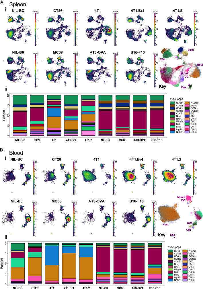 Fig.1 Spleen and blood leukocyte frequencies determined using backbone panel markers.1
Why Choose Us?
Creative Biolabs, a leader in immunological research, offers leukocyte profiling services distinguished by unparalleled expertise, cutting-edge technology, and a commitment to client success. Their highly skilled immunologists and bioinformaticians ensure optimal experimental design and insightful data interpretation. They utilize the latest platforms, including high-parameter flow cytometers, and single-cell RNA sequencing systems, providing comprehensive and accurate data. Creative Biolabs offers customized solutions and adheres to rigorous quality control measures, guaranteeing reliable and reproducible results. Their dedicated bioinformatics team provides in-depth analysis and biological interpretation, translating complex data into actionable insights to accelerate decision-making.
Reach out to discover how our specialized services for the identification, quantification, and comparison of leukocyte populations can enhance your research, clinical diagnostics, or drug development projects.
Frequently Asked Questions
Q1: What types of samples can I submit for leukocyte profiling at Creative Biolabs?
A1: Creative Biolabs processes diverse samples—PBMCs, whole blood, fresh/frozen tissues, dissociated tumors, and biological fluids. Our experts guide optimal collection/preparation to ensure high-quality results for your project.
Q2: Can Creative Biolabs' service help me identify rare or novel leukocyte populations?
A2: Creative Biolabs utilizes Flow Cytometry for high-dimensional immune cell analysis, enabling unbiased discovery and deep phenotyping of rare cell populations—delivering insights beyond conventional methods.
Q3: Can I customize the antibody panels or analysis parameters for my specific research needs with Creative Biolabs?
A3: Creative Biolabs specializes in customized antibody panels and analysis parameters for flow cytometry. We collaborate closely to tailor solutions for your specific research needs. Contact us to discuss your project requirements.
Customer Review
-
Precision in Biomarker Identification
Creative Biolabs' service provided the precision needed to identify subtle shifts in regulatory T cell populations, which we then validated as a key biomarker for therapeutic response in our autoimmune disease model. Their detailed comparative analysis was invaluable. - Lis***a W
-
Overcoming Sample Limitations
We had very limited clinical sample volumes, but Creative Biolabs' optimized protocols for low-input samples allowed us to successfully perform comprehensive leukocyte profiling, yielding critical data we couldn't obtain elsewhere. - Mic***l B
Related Services
To further support your immunological and drug discovery endeavors, Creative Biolabs offers a suite of complementary services that can seamlessly integrate with your leukocyte profiling project:
Chemokine Production Measurement Service
Tregs Migration Assay
At Creative Biolabs, we offer premier Tregs migration assay services. Our services include high-purity isolation of Tregs, fluorescent labeling, and various migration assays such as transwell, real-time tracking, and 3D models. We also provide chemokine and cytokine analysis and detailed data analysis, and reporting.
Learn More →How to Contact Us
Ready to advance your immunological research? Our team of experts at Creative Biolabs is eager to discuss your project and provide a customized solution tailored to your specific needs.
Contact Our Team for More Information and to Discuss Your Project
Reference
- Simon Davis, David A., et al. "Identifying cancer-associated leukocyte profiles using high-resolution flow cytometry screening and machine learning." Frontiers in Immunology 14 (2023): 1211064. Distributed under Open Access license CC BY 4.0, without modification.
 Download our brochure
Loading case studies...
Online Inquiry