
Creative Biolabs is offering the most comprehensive services for antibody development projects. With strict regulation and effective execution, we are dedicated to providing the most valuable solutions to complete your projects.
HighlightsCreative Biolabs is offering the most comprehensive services for antibody development projects. With strict regulation and effective execution, we are dedicated to providing the most valuable solutions to complete your projects.
HighlightsCreative Biolabs has established a broad range of platforms for developing novel antibodies or equivalents. These cutting-edge technologies enable our scientists to meet your demands from different aspects and tailor the most appropriate solution that contributes to the success of your projects.
HighlightsWith deep understanding in antibody-related realms and extensive project experience, Creative Biolabs offers a variety of references to help you learn more about our capacities and achievements, including infographic, flyer, case study, peer-reviewed publications, and all kinds of knowledge that can assist your projects. You are also welcome to contact us directly for more specific solutions.
HighlightsGet a real taste of Creative Biolabs, one of the most professional custom service providers in the world. We are committed to providing highly customized comprehensive solutions with the best quality to advance your projects.
Highlights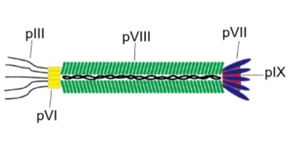
Phage display technology has emerged as a transformative solution, offering a powerful method to link a phenotype (the displayed peptide or protein) to its genotype (the corresponding DNA sequence) through the utilization of bacteriophages as versatile molecular display vehicles. Among the various phage systems, the M13 filamentous phage system is overwhelmingly preferred for its stability, ease of manipulation, and unparalleled capacity for large-scale library screening. The M13 bacteriophage is a non-lytic, single-stranded DNA virus that exclusively infects Escherichia coli strains carrying the F-pilus. Its distinct morphology as a filamentous particle, measuring approximately 6 nm in diameter and 880 nm in length, is structurally defined by its well-characterized, circular, single-stranded genome encapsulated by five primary structural proteins: pIII, pVI, pVII, pVIII, and pIX.
Creative Biolabs is at the forefront of this field, offering comprehensive development and screening services centered on the M13 display system, enabling the isolation of high-affinity peptides, proteins, and antibody fragments from libraries comprising millions of distinct molecular entities. This foundational technology drastically shortens the development cycle for novel binders and reduces production costs by eliminating the need for conventional animal immunization protocols.
Leverage Our End-to-End M13 Display Solutions to Rapidly Advance Your Research Pipeline
Effective phage display hinges on the ability to fuse the heterologous (exogenous) sequence (the peptide, scFv, or Fab) to a phage coat protein without compromising the phage's assembly, stability, or infectivity. The M13 system offers two primary coat proteins, pIII and pVIII, each providing unique strategic advantages based on the size of the insert and the desired display valency.
The pIII protein, essential for phage infection of E. coli, serves as the most common display site for large peptides and complex protein domains, including antibody fragments.
Start Your M13 Display Project Today

Creative Biolabs leverages its deep knowledge and extensive experience to operate a state-of-the-art Phage Display Platform that overcomes the common technical hurdles associated with library construction and screening. Our expertise in manipulating the M13 system ensures that clients benefit from stable, high-titer, and biologically robust phage libraries.
We have refined the genetic engineering techniques necessary for precise control over valency (monovalent or multivalent display), offering unparalleled flexibility in screening strategies tailored to the target antigen's characteristics and the required binder affinity. Our proprietary methods for managing helper phage infection and expression ratios maximize the diversity and quality of the output, directly translating into a higher success rate for identifying lead candidates.
Request a Consultation with Our M13 Specialists

Creative Biolabs offers an end-to-end suite of services built upon the highly efficient M13 Phage Display Platform. Our services are designed to address the entire discovery pipeline, from initial library creation to the isolation of functionally validated binders.
 M13 Phage Display Library Construction Service
We specialize in constructing high-diversity, functional libraries (up to 1011 independent clones), including naïve, synthetic, and immune libraries, ensuring comprehensive coverage of sequence space for optimal target binding.
Phage Display Library Screening Service (Biopanning)
Utilizing tailored screening protocols, we perform rigorous biopanning against various targets (proteins, cells, tissues, small molecules) to enrich for high-affinity clones, employing sophisticated techniques for counter-selection and elution to maximize specificity.
Phage Display-based Monoclonal Antibody Discovery Service
This is a core strength of our platform, enabling the rapid isolation of fully human or humanized antibody fragments (scFv, Fab) against almost all antigens, circumventing the need for in vivo immunization and offering a faster route to therapeutic development.
Phage Display-based Peptide Discovery Service
We excel at identifying short, high-affinity peptide ligands that can serve as targeting moieties, receptor agonists/antagonists, or structural probes, typically displayed on the pVIII coat protein.
Phage Display-based Stable Binder Discovery Service
We employ specialized selection strategies to isolate binders with enhanced thermal stability, resistance to proteases, or favorable properties for long-term storage and manufacturing.
Phage Display-based pH-Sensitive Binder Discovery Service
Crucial for drug delivery applications, this service focuses on isolating binders that exhibit altered binding affinity contingent upon changes in pH, enabling targeted release in acidic microenvironments (e.g., tumors or endosomes).
Phage Display-based Internalizing Antibody Discovery
Through our phage display platform, we pinpoint antibodies with the rare ability to initiate receptor-mediated internalization—unlocking precise transport of drugs or biological payloads directly into target cells.
Transform Your Molecule Screening Strategy with Our High-Throughput M13 Display Services
Discuss Your Screening Needs with Our Experts
All listed services and products are For Research Use Only. Do Not use in any diagnostic or therapeutic applications.








