
Creative Biolabs is offering the most comprehensive services for antibody development projects. With strict regulation and effective execution, we are dedicated to providing the most valuable solutions to complete your projects.
HighlightsCreative Biolabs is offering the most comprehensive services for antibody development projects. With strict regulation and effective execution, we are dedicated to providing the most valuable solutions to complete your projects.
HighlightsCreative Biolabs has established a broad range of platforms for developing novel antibodies or equivalents. These cutting-edge technologies enable our scientists to meet your demands from different aspects and tailor the most appropriate solution that contributes to the success of your projects.
HighlightsWith deep understanding in antibody-related realms and extensive project experience, Creative Biolabs offers a variety of references to help you learn more about our capacities and achievements, including infographic, flyer, case study, peer-reviewed publications, and all kinds of knowledge that can assist your projects. You are also welcome to contact us directly for more specific solutions.
HighlightsGet a real taste of Creative Biolabs, one of the most professional custom service providers in the world. We are committed to providing highly customized comprehensive solutions with the best quality to advance your projects.
Highlights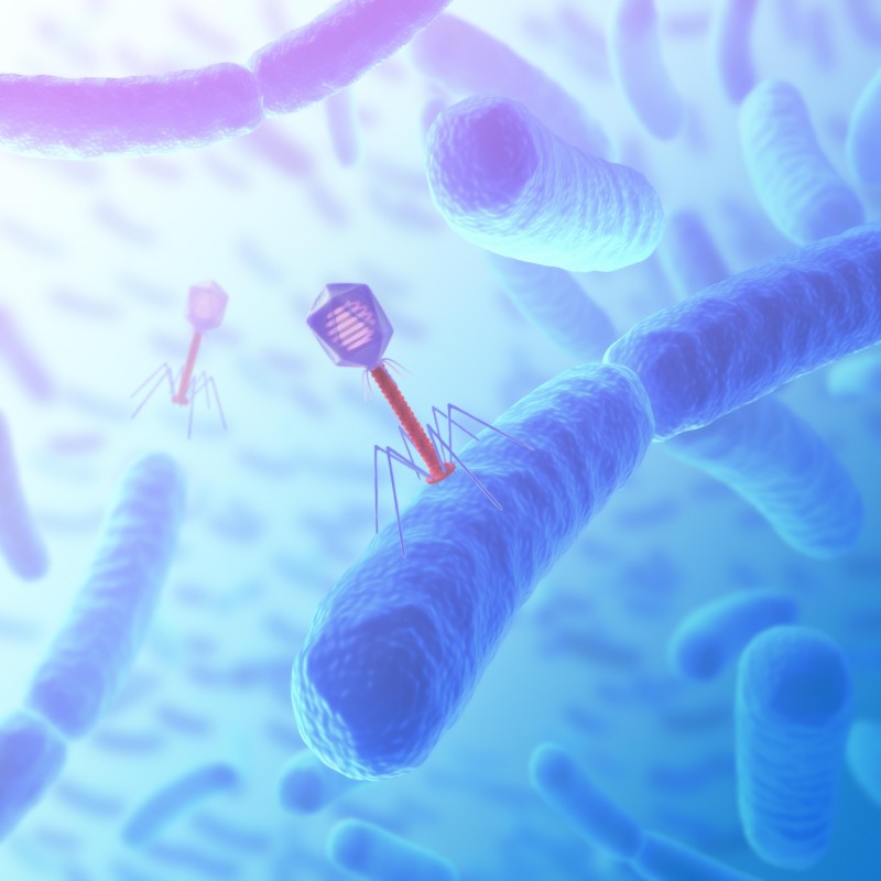
Phage display is an elegant molecular biology technique that physically links an exogenous polypeptide or protein to the genetic information (DNA) encoding it. This is achieved by inserting the gene segment of interest into the appropriate position within the structural gene of a phage capsid protein. The insertion must be in the correct reading frame and must not compromise the essential function of the capsid protein, which is critical for phage assembly and infectivity.
Upon expression, the exogenous gene is translated along with the capsid protein, forming a fusion protein. This fusion protein is subsequently incorporated into the structure of the nascent bacteriophage, resulting in the display of the foreign peptide or protein on the phage surface. This linkage of phenotype (the displayed protein) and genotype (the packaged DNA) is the fundamental principle that makes phage display a high-throughput screening platform. It allows researchers to select for phages displaying a desired binding characteristic—such as high affinity for a target molecule—and then readily identify the sequence encoding that binder simply by sequencing the DNA. The entire process forms a cyclical selection and amplification strategy.
Let Our Phage Display Experts Guide Your Discovery. Request a Consultation.
The effectiveness and application range of phage display are critically dependent on the choice of the phage vector. Different phages offer distinct advantages concerning display capacity, copy number, size of the insert, and stability. Creative Biolabs expertly utilizes and offers four primary bacteriophage systems: M13, T4, T7, and Lambda phages, each optimized for specific experimental requirements.
| Phage System | Genome & Life Cycle | Display Site / Mechanism | Copy Number | Best For | Key Advantages |
| M13 Phage Display | ssDNA circular virus (6.4 kb), non-lytic secretion — continuous production | PIII (low-copy) or PVIII (high-copy) coat proteins |
• PIII: 3-5 copies • PVIII: 100s–1000s copies |
• Antibody fragment libraries (scFv, Fab) • Peptide libraries |
• Most mature & widely used system • Tunable display valency • Highly robust, even explored in toxicology |
| T4 Phage Display | dsDNA linear virus, lytic; large genome | Fusion to SOC (C-terminus) and/or HOC (N-terminus) | Extremely high — simultaneous SOC + HOC display |
• Large / folded proteins • Toxic proteins |
• Non-essential SOC/HOC ensures high stability • High avidity binding • Excellent for difficult proteins |
| T7 Phage Display | dsDNA linear virus, lytic | Fusion to capsid protein 10B |
• High copy: small peptides ≤50 aa • Medium/low copy: large proteins ≤1200 aa |
• Both peptides & large proteins • Affinity/size flexible projects |
• Adjustable copy number • Efficient display of large proteins • Ideal for broad screening applications |
| Lambda Phage Display | dsDNA linear virus (48.5 kb) with cohesive ends; temperate | Head proteins D and V | Medium to high |
• Structural / large functional proteins (>100 kDa) • Toxic proteins |
• Entire assembly in cytoplasm — no secretion limits • Best for difficult-to-fold or toxic proteins |
Tell Us Your Target, We'll Design the Optimal Phage Display Strategy for You
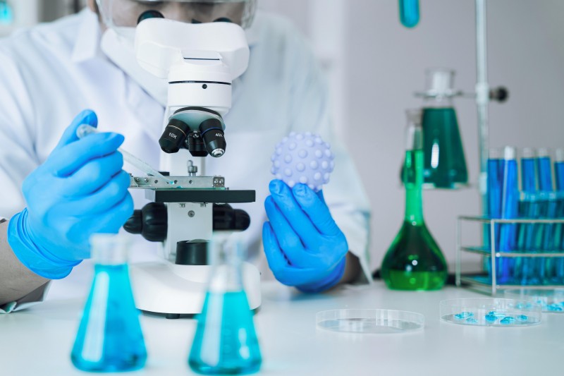
Creative Biolabs leverages a wealth of knowledge and experience to deliver a high-performance Phage Display Platform that surpasses industry standards. We recognize that the success of a phage display project hinges on meticulous library construction and optimized selection protocols. Our platform is characterized by:

Our commitment to precision, high throughput, and robust quality control ensures that researchers receive the most reliable and advanced tools to accelerate their discovery programs.
Start Your Antibody or Peptide Screening with Confidence. Speak With a Scientist.

Creative Biolabs offers a comprehensive suite of services built around our core Phage Display Platform. These services are designed to facilitate the discovery and development of novel therapeutic and diagnostic agents.
We provide end-to-end solutions for Phage Display based Binder Discovery, encompassing all critical stages of the selection process:
Generating customized high-quality naive, semi-synthetic, or immune libraries (e.g., VHH, scFv, Fab libraries) tailored to specific research needs.
Employing specialized biopanning protocols (e.g., solid-phase, solution-phase, in vivo screening) to efficiently isolate target-specific binders from complex libraries.
Rapid and reliable isolation of high-affinity monoclonal antibody fragments (scFv, Fab) against virtually any antigen, a crucial first step for therapeutic antibody development.
Identification of novel peptide ligands with high affinity and specificity, which can serve as research tools, diagnostic agents, or drug candidates.
Focused screening to identify binders that exhibit enhanced stability under physiological or storage conditions, improving their utility and manufacturability.
Specialized selection protocols to isolate binders that display pH-dependent binding characteristics, which are particularly valuable for developing agents with enhanced efficacy in acidic tumor microenvironments or for improved recycling in endosomes (resulting in superior half-life).
Discovering antibodies that activate receptor-mediated entry, ensuring that attached therapeutic molecules are efficiently transported inside the target cells.
Ready to Launch Your Phage Display Project? Request a Quote.
Your Success Starts with the Right Platform. Talk to Our Project Managers.
All listed services and products are For Research Use Only. Do Not use in any diagnostic or therapeutic applications.








