
Creative Biolabs is offering the most comprehensive services for antibody development projects. With strict regulation and effective execution, we are dedicated to providing the most valuable solutions to complete your projects.
HighlightsCreative Biolabs is offering the most comprehensive services for antibody development projects. With strict regulation and effective execution, we are dedicated to providing the most valuable solutions to complete your projects.
HighlightsCreative Biolabs has established a broad range of platforms for developing novel antibodies or equivalents. These cutting-edge technologies enable our scientists to meet your demands from different aspects and tailor the most appropriate solution that contributes to the success of your projects.
HighlightsWith deep understanding in antibody-related realms and extensive project experience, Creative Biolabs offers a variety of references to help you learn more about our capacities and achievements, including infographic, flyer, case study, peer-reviewed publications, and all kinds of knowledge that can assist your projects. You are also welcome to contact us directly for more specific solutions.
HighlightsGet a real taste of Creative Biolabs, one of the most professional custom service providers in the world. We are committed to providing highly customized comprehensive solutions with the best quality to advance your projects.
HighlightsPhage display kits represent integrated reagent systems that are pre-assembled to enable laboratories to adopt phage display technology easily. The kits simplify complex phage display library construction and screening through pre-made components such as engineered phage vectors and optimized bacterial strains and user protocols. The use of phage display kits helps speed up experimental processes by lowering technical obstacles which results in reduced variability and improved reproducibility among different laboratories.
These kits provide researchers with a quick method to identify target-specific ligands that demonstrate both high affinity and specificity. Phage display kits enable users to concentrate on experimental design and downstream analysis because they do not require specialized molecular cloning and propagation techniques like traditional library construction does.
These kits serve a critical role in:
Phage display kits provide universal access to advanced molecular selection instruments which empower both novice researchers and experienced professionals to perform significant research activities quickly while saving money and scaling operations.
Phage display kits exist in multiple specialized formats designed to suit particular molecular targets and expression systems alongside research goals. Researchers must evaluate multiple criteria including target molecule characteristics, binding ligand format and downstream application goals when choosing a phage display kit. The available kits are divided into multiple main categories which provide specific biological functions and technical benefits.
Phage display peptide library kits enable researchers to showcase extensive collections of random or semi-random peptides that span 7 to 12 amino acids in length. Filamentous phages such as M13 host these libraries which are genetically attached to coat proteins like pIII or pVIII and then expressed in E. coli host strains. The random peptide sequences and their extensive diversity enable unbiased testing across numerous targets. Peptide libraries exist as linear structures or as constrained cyclic forms with disulfide bridges which demonstrate increased binding stability and specificity.
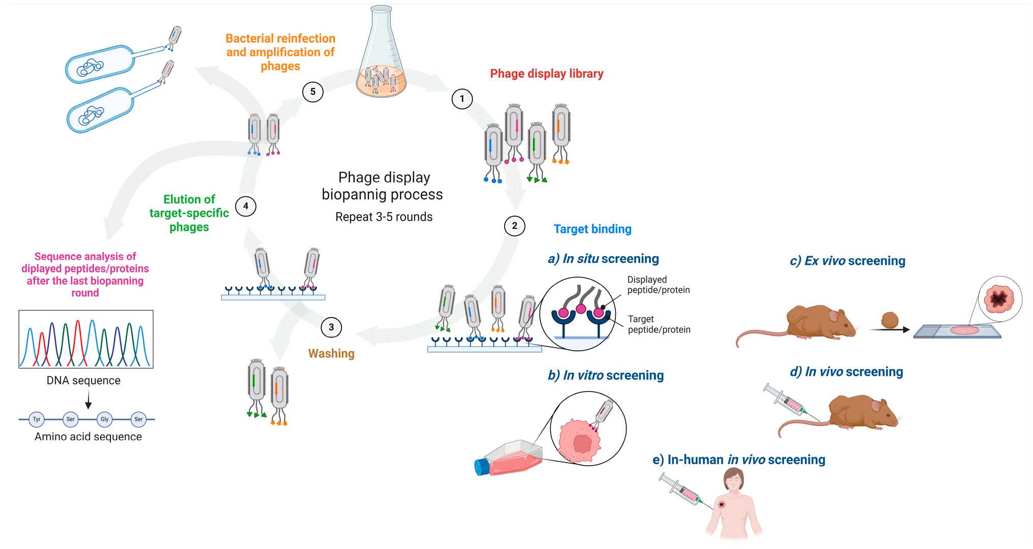 Fig. 1 Schematic of an affinity-driven process in which phage-displayed libraries are screened against a variety of targets and target-specific phage-displayed (poly)peptides (e.g., novel cancer ligands) are subsequently identified.1, 3
Premade Peptide Library Licensing from Creative Biolabs:
Empowered by extensive experience and well-established phage display technique platform, Creative Biolabs now brings out a comprehensive list of premade peptide libraries for various research programs. Of note, we carry 2 good linear peptide libraries (16-mer, and 20-mer), which are based on the same true phage system as NEB Ph.D. peptide libraries.
In addition, we also carry following premade peptide libraries for your selection:
| Peptide Library ID | Phage Display | Library Format | Library Size |
| TriCo-C9L | pIII-fusion, Phagemid Phage Display | Cyclic 9-mer peptide library | 6.8×10¹⁰ |
| Protease-10A | pIII-fusion, Phagemid Phage Display | HiAffi tagged 10 mer peptide library | 1.10×10¹⁰ |
Phage display antibody library kits consist of pre-made libraries with antibody fragments like scFv or Fab displayed on bacteriophage surfaces. The antibodies found in these libraries originate from human, mouse, or synthetic immunoglobulin gene collections which allow users to select antigen-specific antibodies in vitro without immunization.
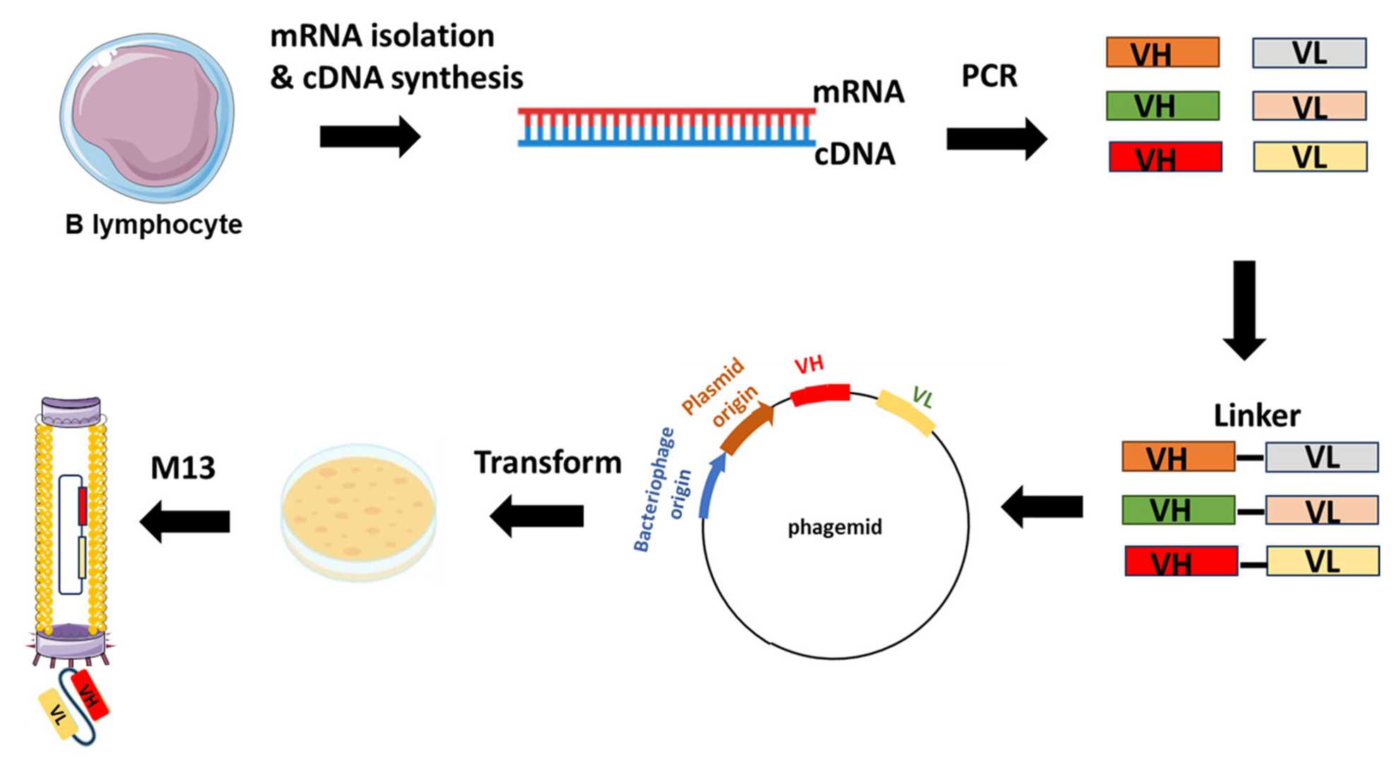 Fig. 2 Example of scFv-phage display library construction.2, 3
scFv libraries feature variable domains VH and VL which are joined together by a small peptide spacer through genetic fusion. The small size of these molecules allows for simpler expression and handling which makes them perfect for high-throughput selection processes.
| Libraries | Library ID | Display Technology | Library Format | Species | Library Size |
| Phage display human scFv libraries | HuScL-2 | Phage Display | Semi-synthetic scFv | Human | 1.42×10⁹ |
| HuScL-4 | Phage Display | Naïve scFv | Human | 2.0×10⁹ | |
| HuScL-6 | Phage Display | Naïve scFv | Human | 1.0×10¹¹ | |
| HuScL-7 | Phage Display | Naïve scFv | Human | 1.1×10¹⁰ | |
| HuScL-2S | Phage Display | scFv | Human | 2.36×10¹⁰ |
Fab fragments contain the constant domains CH1 and CL in addition to VH and VL which result in enhanced stability and improved biophysical characteristics. The libraries serve as standard tools for both therapeutic development and clinical-grade antibody engineering.
| Libraries | Library ID | Display Technology | Library Format | Species | Library Size |
| Phage display human Fab libraries | HuFabL-4 | Phage Display | Naïve Fab | Human | 3.0×10¹⁰ |
| HuFabL-5 | Phage Display | Naïve Fab | Human | 1.1×10¹⁰ | |
| HuFabssL-1 | Phage Display | Naïve & synthetic Fab | Human | 1.8×10¹⁰ |
In addition to filamentous M13 phage systems, T4 and T7 bacteriophage platforms offer alternative display environments. Unlike M13, which relies on secretion through the bacterial membrane, T4 and T7 are lytic phages that allow expression of larger and more complex proteins within the bacterial cytoplasm, enhancing folding and yield.
Phagemid vectors or full phage genomes are engineered to include:
The host bacterial strain is critical for efficient phage propagation, protein folding, and genetic stability. Strains must be compatible with the vector system and optimized for phage assembly.
Key Considerations in Host Selection:
| Strain | Genotype | Purpose |
| E. coli ER2738 | F', lacZΔM15, high transformation rate | Standard for M13 amplification and selection |
| E. coli TG1 | supE, thi, lacZΔM15 | Versatile strain for antibody library screening |
| E. coli BL21 (DE3) | T7 polymerase, protease-deficient | Used with T7-based phage display systems |
| E. coli XL1-Blue | recA1, endA1, lacIq | High-fidelity cloning and vector propagation |
Phage display accelerates ligand identification, especially for kinases, GPCRs, and cytokines. Hits from kits can be optimized through affinity maturation.
Fine mapping of linear or conformational epitopes supports monoclonal antibody development and disease-specific biomarker detection.
Protein-binding peptides or antibody fragments selected via phage display can map interaction domains or screen inhibitors for disrupting interfaces.
Commercially optimized kits can implement phage display technology to provide a flexible and effective molecular discovery platform. Phage display technology dominates academic and industrial research because it can analyze large molecular libraries under specific conditions for antibody engineering and peptide drug screening. Just as all technologies contain built-in disadvantages. The ability to design efficient and goal-oriented experiments depends on understanding both the strengths and limitations of the technology.
Phage display kits offer a multitude of technical, operational, and strategic advantages, particularly when compared to traditional molecular screening platforms.
| Benefit | Description | Research/Industrial Value |
| Rapid and Scalable Discovery | Enables screening of up to 10⁹–10¹¹ variants within days using automated biopanning cycles | Accelerates lead identification, ideal for time-sensitive projects |
| Genotype–Phenotype Linkage | Each phage links displayed protein to its encoding DNA, simplifying downstream identification | Facilitates efficient hit validation and cloning |
| Ready-to-Use Format | Kits include optimized vectors, host strains, and reagents | Minimizes preparation time, reduces experimental error |
| Cost Efficiency | Uses inexpensive bacterial systems without need for animals or advanced equipment | Makes discovery accessible to both startups and academic labs |
| Versatility of Applications | Supports peptide, antibody fragment, and protein display across diverse targets | Applicable to drug discovery, diagnostics, epitope mapping, and vaccine research |
| Customizable Display Systems | Available in M13, T7, or T4 formats with different display valency and protein sizes | Tailorable to experimental goals (e.g., small peptides vs. full-length proteins) |
| Seamless Downstream Engineering | Sequences from selected phages can be cloned into expression or therapeutic formats | Enables affinity maturation, humanization, or structural conversion for development |
| High Library Diversity | Commercial kits offer libraries with up to 10¹¹ unique variants | Maximizes likelihood of identifying high-affinity, target-specific binders |
| Standardization and Reproducibility | Pre-validated kits minimize variability and enhance result comparability | Important for regulated environments, such as biotech R&D and CRO pipelines |
Despite its broad utility, phage display is not without limitations. These challenges must be considered to optimize screening conditions and ensure biological relevance of the identified binders.
| Challenge | Root Cause / Impact | Recommended Solutions / Best Practices |
| Protein folding limitations | Misfolded proteins due to lack of PTMs in E. coli | Use Fab or scFv instead of full-length antibodies; switch to T7 display system; validate folding via ELISA or Western blot |
| Display bias during panning | Preferential amplification of toxic or overexpressed clones | Reduce amplification cycles; include negative selection steps; perform next-gen sequencing (NGS) post-panning |
| Conformational epitope inaccessibility | Linear peptides cannot mimic native protein folding | Use cyclic peptide libraries or full Fab libraries; incorporate structural modeling of target antigen |
| Non-specific binding (high background) | Interaction with plastic surfaces or blocking reagents | Optimize blocking buffers (e.g., 5% milk vs. BSA); add stringent wash steps; include negative control targets |
| Loss of rare binders in amplification | Overgrowth of fast-replicating, low-affinity phages | Use low-MOI amplification; minimize outgrowth time; analyze diversity with Sanger or NGS across rounds |
| Host strain limitations | Incompatibility of certain vectors or low expression efficiency | Use phage system-compatible strains (e.g., ER2738 for M13, BL21(DE3) for T7); test expression before panning |
| Discrepancy between in vitro/in vivo | Differences in buffer, folding, expression systems | Validate hits in mammalian cells; clone into full-length IgG or expression vectors for functional assays |
| Translational gap for therapeutics | Non-human or immunogenic sequences may not be suitable for clinical use | Humanize selected antibodies; run immunogenicity prediction tools; perform affinity maturation |
Over years, Creative Biolabs has established a comprehensive list of libraries for de novo discovery of therapeutically relevant antibodies and peptides. If none of the premade libraries meets your requirement, you are welcome to contact us, and we will be more than happy to create a custom library for you.
Learn more about Creative Biolabs premade phage display library ready-to-use kits and premade phage display services:
All listed services and products are For Research Use Only. Do Not use in any diagnostic or therapeutic applications.








