
Creative Biolabs is offering the most comprehensive services for antibody development projects. With strict regulation and effective execution, we are dedicated to providing the most valuable solutions to complete your projects.
HighlightsCreative Biolabs is offering the most comprehensive services for antibody development projects. With strict regulation and effective execution, we are dedicated to providing the most valuable solutions to complete your projects.
HighlightsCreative Biolabs has established a broad range of platforms for developing novel antibodies or equivalents. These cutting-edge technologies enable our scientists to meet your demands from different aspects and tailor the most appropriate solution that contributes to the success of your projects.
HighlightsWith deep understanding in antibody-related realms and extensive project experience, Creative Biolabs offers a variety of references to help you learn more about our capacities and achievements, including infographic, flyer, case study, peer-reviewed publications, and all kinds of knowledge that can assist your projects. You are also welcome to contact us directly for more specific solutions.
HighlightsGet a real taste of Creative Biolabs, one of the most professional custom service providers in the world. We are committed to providing highly customized comprehensive solutions with the best quality to advance your projects.
HighlightsPlatelet-derived growth factor receptors (PDGFRs) belong to the receptor tyrosine kinase (RTK) family and encompass two structurally related subtypes, PDGFRα and PDGFRβ, encoded by the PDGFRA gene on chromosome 4q12 and the PDGFRB gene on chromosome 5q33, respectively. These cell-surface receptors play indispensable roles in the regulation of fundamental cellular processes, including proliferation, migration, survival, and differentiation. Through the binding of PDGF ligands, PDGFRs govern critical physiological events such as embryonic development, angiogenesis, and wound healing.
Dysregulation of PDGFR signaling is intimately linked to a spectrum of human pathologies. Aberrant activation, whether through overexpression, gene fusion, or activating mutations, drives tumorigenesis in numerous malignancies, promotes the progression of fibrotic diseases, and contributes to atherosclerosis. Consequently, PDGFRs have emerged as high-value targets in both basic research and therapeutic development. High-quality anti-PDGFR antibodies serve as essential tools for dissecting the biological functions of PDGFRs, decoding the underlying signaling networks, and enabling the design of targeted intervention strategies, including Creative Biolabs' custom agonistic antibody development service and agonistic antibody therapy.
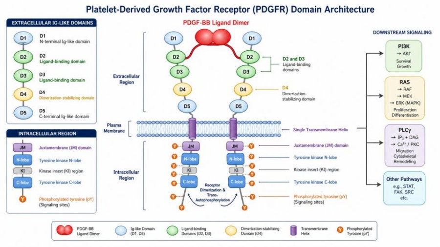
The domain architecture of PDGFR is characteristic of the RTK family, comprising an extracellular ligand-binding domain, a single transmembrane helix, and an intracellular region that houses a juxtamembrane domain and a split tyrosine kinase domain with a kinase insert region. The extracellular portion is organized into five immunoglobulin-like (Ig-like) domains, with the first three domains responsible for ligand recognition and dimerization specificity.
Mechanistically, engagement of cognate PDGF ligands induces receptor dimerization, which brings the intracellular kinase domains into close proximity, triggering trans-autophosphorylation of specific tyrosine residues. These phosphorylated sites serve as docking stations for downstream signaling adaptors, initiating cascades including PI3K/AKT, RAS-mitogen-activated protein kinase (MAPK), and PLCγ pathways. While PDGFRα and PDGFRβ share a conserved overall topology, they exhibit distinct ligand-binding profiles, tissue distributions, and signaling outputs. PDGFRα binds PDGF-AA, PDGF-BB, and PDGF-CC, whereas PDGFRβ is predominantly activated by PDGF-BB and PDGF-DD. These differences necessitate a nuanced antibody design strategy. Anti-PDGFR antibodies can be developed to target the extracellular domain to block ligand binding, or to recognize specific phosphorylation states of the intracellular domain to monitor receptor activation, thereby serving as either functional antagonists or sensitive detection reagents.
Anti-PDGFR antibodies have become indispensable in multiple biomedical research domains. Key application scenarios include:
PDGFR is expressed on tumor-associated fibroblasts, pericytes, and on tumor cells themselves. It actively participates in tumor angiogenesis, interstitial fluid pressure regulation, and immune evasion. Anti-PDGFR antibodies enable immunohistochemistry (IHC), immunofluorescence (IF), and flow cytometric analysis to delineate the spatial distribution and expression levels of PDGFR within complex tumor tissues.
Specific anti-PDGFR antibodies are deployed in Western blot, immunoprecipitation, and kinase activity assays to decipher the cross-talk between the PDGF/PDGFR axis and canonical pathways such as PI3K/AKT and RAS-mitogen-activated protein kinase (MAPK). Phosphorylation-site-specific antibodies further allow precise interrogation of activation dynamics.
PDGFR signaling is a central driver of organ fibrosis, including pulmonary fibrosis, liver fibrosis, and renal fibrosis. In these models, anti-PDGFR antibodies function as blocking tools to attenuate pro-fibrotic responses or as sensitive detection reagents to monitor disease progression and therapeutic efficacy.
Blocking anti-PDGFR antibodies can serve as lead molecules for the development of therapeutic modalities against PDGFR-driven tumors or fibrotic disorders. They represent fundamental building blocks for advanced formats such as antibody-drug conjugates (ADCs) and bispecific antibodies, enabling selective targeting of pathological cells.
Creative Biolabs has established an integrated, technology-diverse antibody discovery platform that has successfully empowered numerous projects targeting receptor tyrosine kinases, including PDGFRα and PDGFRβ. Our platform is architected to address the most challenging targets with flexibility and specificity.
 Multiple Display Technologies
We deploy phage display, yeast display, and mammalian cell display systems. This versatility allows us to select the optimal screening environment based on target conformation, post-translational modifications, and desired antibody format.
Diversified Immunization Strategies
Our offerings encompass protein immunization, DNA immunization, cell-based immunization, and fully human transgenic animal immunization. This portfolio ensures access to a broad repertoire of antibody candidates from various host species.
High-Throughput Screening and Validation
A tiered validation cascade integrates surface plasmon resonance (SPR), ELISA, FACS, and cell-based functional assays. This rigorous pipeline confirms the affinity, specificity, and biological activity of every candidate antibody.
Advanced Antibody Engineering
Downstream optimization services include antibody humanization, affinity maturation, and Fc engineering, transforming primary hits into development-ready leads.

Driven by science and extensive experience, Creative Biolabs delivers custom anti-PDGFR antibodies with high specificity, sensitivity, and lot-to-lot consistency. Our development process starts from rigorous antigen design and host immunization, followed by tailored antibody selection. For polyclonal antibodies, we purify the reactive serum; for monoclonal antibodies, we apply advanced hybridoma technology with high-throughput screening. Whether your goal is a research tool or a therapeutic lead, our integrated service portfolio covers every stage of the project:
From antigen design to lead candidate identification.
High-throughput screening of custom libraries against PDGFR.
Traditional hybridoma platform integrated with high-throughput screening.
One-stop recombinant antibody production from gene synthesis to purified protein.
Development of robust detection methods based on anti-PDGFR antibodies.
All listed services and products are For Research Use Only. Do Not use in any diagnostic or therapeutic applications.








