Based on the target organ, we select the appropriate ECM scaffold and cell types to represent the tissue-specific TME.
Angiogenesis-Tumor Microenvironment Interaction Modeling Service
Angiogenesis-tumor microenvironment interaction modeling service at Creative Biolabs helps you optimize therapeutic efficacy and predict response through advanced 3D microfluidic vascularized chips and multiscale computational systems biology. We bridge the gap between static in vitro assays and complex human physiology, ensuring your lead compounds are tested in a realistic, perfusion-capable environment that mimics the chaotic vascular nature of malignancies. By integrating precise biomechanical cues and cellular cross-talk, we empower your team to de-risk development and gain deep insights into the spatial distribution of your candidates within the specialized tumor landscape.
Overview What We Can Offer Workflow Required Materials Application Highlights Customer Reviews FAQs Related Services
Overview
Tumor progression is a multi-scale process where hypoxia triggers a cascade of pro-angiogenic signals, resulting in a dysfunctional vascular network. Research indicates that the tumor microenvironment (TME) significantly influences drug resistance through physical barriers like high interstitial fluid pressure and biological shifts like the epithelial-mesenchymal transition (EndMT). Advanced studies demonstrate that integrating agent-based modeling with 3D bioprinting allows for a precise simulation of these dynamics. Sophisticated modeling of these interactions is essential for developing effective anti-angiogenic therapies and improving the delivery of chemotherapeutic and immunotherapeutic agents within experimental frameworks.
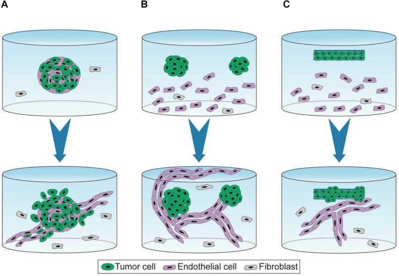 Fig.1 3D experimental platforms designed to study angiogenic capillary formation and tumor growth dynamics.1
We employ a multi-layered strategy to capture the bidirectional communication between malignant cells and the surrounding vasculature. By utilizing human primary cells, we replicate the specific metabolic and mechanical cues of the TME. Our approach focuses on modeling vascular normalization—the process of temporary vessel stabilization—to identify the optimal window for drug administration. Furthermore, we integrate spatial transcriptomics data with our computational models to map the heterogeneity of endothelial cells, ensuring that the simulated environment reflects the true complexity of patient-specific tumor landscapes.
Empower Your Research with Creative Biolabs — Begin Your Expert Consultation Today
What We Can Offer
Vascularized Tumor-on-a-Chip Platforms
We provide customized 3D microfluidic systems that feature perfusable endothelial lumens alongside stromal and immune compartments, allowing for the study of real-time extravasation.
Multiscale Computational Modeling
Our computational services include the development of hybrid discrete-continuous models that simulate oxygen gradients, nutrient diffusion, and drug transport kinetics across dense tumor tissue.
High-Resolution Morphometric Analysis
We deliver quantitative data on vessel morphology, including branching density, tortuosity, and flow velocity, providing a clear picture of how your compounds alter vascular architecture.
Tissue-Specific ECM Customization
Our team engineers organ-specific extracellular matrices (e.g., bone, brain, or liver) to mimic the distinct mechanical stiffness and biochemical signaling of different secondary sites.
Immune Cell Infiltration Assays
We offer specialized modules to monitor the migration of T-cells and myeloid cells through the vascular wall into the tumor core under various experimental treatment conditions.
Workflow
Model Selection and Design
Cell Sourcing and Preparation
Creative Biolabs utilizes high-quality primary human endothelial cells, fibroblasts, and tumor cell lines, ensuring all biological components are validated for phenotypic consistency.
Microfluidic Chip Assembly
We construct the vascularized environment using precision 3D bioprinting or microfluidic fabrication, establishing a stable, perfusable network within the 3D matrix.
Experimental Execution
We introduce your therapeutic agents into the system under controlled flow conditions, monitoring real-time interactions using confocal or multi-photon microscopy.
Data Acquisition and Computational Integration
Raw imaging data is integrated into our computational models to quantify parameters like vessel permeability and drug distribution volumes.
Final Reporting and Insight
You receive a detailed technical report containing statistical analyses, high-resolution imagery, and expert interpretations to guide your next development phase.
Required Starting Materials
- Target Profile: Detailed information on the drug's mechanism of action (e.g., small molecule, monoclonal antibody, or cell therapy).
- Cell Lines or Samples: Specific tumor cell lines of interest or patient-derived samples if personalized modeling is required.
- Benchmarking Data: Any existing in vitro data that can assist in calibrating the model for your specific compound.
Application
Anti-Angiogenic Compound Screening
Our platforms serve as a high-fidelity filter to evaluate the potency of compounds designed to inhibit endothelial sprouting or promote vascular pruning in a human-relevant context.
Drug Delivery Optimization
We model the impact of interstitial fluid pressure and vascular leakiness to determine how specific formulations can better penetrate the dense stromal barriers of solid tumors.
Combination Therapy Evaluation
Researchers use our models to identify the ideal timing for administering secondary drugs following a vascular-normalizing agent to maximize intra-tumoral concentration.
Mechanistic Studies of Resistance
Our services allow for the detailed investigation of non-angiogenic growth patterns, such as vessel co-option and vasculogenic mimicry, which often drive treatment failure.
Highlights
Physiological Flow Simulation
Our systems go beyond static culture by recreating the precise shear stress and interstitial pressure that dictate drug delivery within a living organism. By accurately simulating these fluid dynamics, we help you understand the physical constraints that often prevent even the most potent molecules from reaching the tumor center in a laboratory setting.
Human-Centric Systems
We eliminate the noise of species-specific bias by using entirely human-derived cellular components and recombinant matrices. This focus ensures that the signaling pathways and molecular interactions observed in our models are directly relevant to human biology, providing a superior level of data confidence compared to traditional rodent-based assays.

Multiscale Precision Modeling
Our unique value lies in the seamless integration of physical 3D models with powerful in silico simulations for comprehensive data triangulation. This dual approach allows us to quantify microscopic cellular events while simultaneously predicting macroscopic tumor growth trends, offering a holistic view of the vascular-tumor interaction.
High-Throughput Scalability
Our platforms are meticulously designed for scalability, allowing for the screening of multiple compounds or varying concentrations simultaneously without compromising biological complexity. This efficiency enables your team to accelerate the lead optimization phase by generating robust, statistically significant datasets in a fraction of the time required by legacy methods.
Experience the Creative Biolabs Difference — Contact Us for a Tailored Quote
Customer Reviews
- "[Improved Infiltration Metrics] Using Creative Biolabs' angiogenesis-tumor microenvironment interaction modeling service in our research has significantly improved our ability to visualize T-cell extravasation. The perfusable chip design allowed us to see exactly why our lead antibody was failing to penetrate the stromal barrier, a detail we missed in other models." 4 months ago, Dr. H***en L.
- "[Optimized Dosing Windows] Using Creative Biolabs' angiogenesis-tumor microenvironment interaction modeling service in our research has significantly facilitated our discovery of the vascular normalization window. The computational integration was spot-on, matching our later pilot study results perfectly while saving us months of trial and error." 7 months ago, Sarah M***ez.
FAQs
How does your 3D model compare to traditional 2D tube formation assays?
While 2D assays are useful for basic screening, they lack the flow dynamics, ECM stiffness, and 3D architecture of a real tumor. Our service provides a 3D environment with controlled perfusion, which is essential for studying drug transport and the physical barriers present in the tumor microenvironment.
What parameters do you use to measure "vascular normalization"?
We track changes in vessel diameter, basement membrane coverage, pericyte recruitment, and reductions in permeability. These quantitative metrics help determine if a therapy is making the tumor vasculature more structured and thus more receptive to drug delivery.
Do your models account for the metabolic state of the tumor, such as hypoxia?
Yes, our systems are designed to create and monitor oxygen gradients. We use oxygen-sensitive probes to ensure the "hypoxic core" of the simulated tumor mimics the low-oxygen conditions found in solid malignancies, which is a major driver of angiogenesis.
Related Services
Endothelial Tube Formation & Sprouting Analysis Service
This service quantitatively evaluates endothelial tube formation and sprouting in vitro by measuring parameters such as tube length, branching, and network complexity. It enables efficient assessment of angiogenic potential in response to genes, biomolecules, or test compounds.
Learn More →Endothelial Cell Migration & Invasion Analysis Service
This service provides quantitative analysis of endothelial cell migration and invasion using established in vitro models. Key endpoints include migration distance and invasion capacity, supporting studies of angiogenesis and endothelial function regulation.
Learn More →Creative Biolabs offers a sophisticated, multi-scalar approach to Angiogenesis-Tumor Microenvironment Interaction Modeling. By combining state-of-the-art microfluidics with robust computational analysis, we provide a predictive platform that significantly enhances the understanding of vascular dynamics in oncology research. Our team of experts is dedicated to helping you overcome the hurdles of the TME, ensuring your research leads to a deeper understanding of therapeutic delivery and vascular response.
Contact Our Team to Discuss Your Angiogenesis and Tumor Microenvironment Modeling Project.
Reference
- Brassard-Jollive, Noémie et al. "In vitro 3D Systems to Model Tumor Angiogenesis and Interactions With Stromal Cells." Frontiers in cell and developmental biology vol. 8 594903. 5 Nov. 2020. Distributed under Open Access license CC BY 4.0, without modification. https://doi.org/10.3389/fcell.2020.594903
 Download our brochure
Loading case studies...
Online Inquiry