We meet with your team to define the research goals, select the optimal cell lines (e.g., HUVEC, HMEC-1), and determine whether a 2D tube formation or 3D sprouting model is most appropriate.
Endothelial Tube Formation & Sprouting Analysis Service
Our endothelial tube formation & sprouting analysis service at Creative Biolabs helps you obtain precise, high-throughput vascular data and accelerate drug discovery through advanced 3D spheroid technologies, basement membrane modeling, and AI-driven automated image analysis. We provide translatable insights that bridge the gap between in vitro screening and in vivo success.
Overview What We Can Offer Workflow Required Materials Highlights Publication Customer Reviews FAQs Related Services
Overview
The study of angiogenesis is pivotal for therapeutic advancement in oncology, wound healing, and cardiovascular disease. Literature highlights that while 2D tube formation on basement membrane extracts provides rapid screening, it primarily models endothelial reorganization. In contrast, 3D sprouting assays using spheroids better mimic the tip-and-stalk cell hierarchy and matrix invasion seen in vivo. Studies published in journals like Nature and Scientific Reports emphasize that quantifying parameters such as cumulative sprout length and junction density is essential for evaluating drug efficacy. Creative Biolabs integrates these methodologies to provide a validated, high-credibility platform for vascular research.
We employ a multi-tiered strategy to ensure the highest biological relevance for your project. Our approach begins with selecting the most appropriate cell type, typically low-passage Human Umbilical Vein Endothelial Cells (HUVECs) or organ-specific microvascular cells, to match your target tissue. We utilize growth-factor-reduced (GFR) matrices to eliminate background interference when testing pro-angiogenic compounds. For complex drug mechanisms, we implement 3D fibrin or collagen-embedded spheroid assays, which allow for the observation of true invasive sprouting and lumen formation. This strategic combination ensures that both rapid reorganization and invasive morphogenesis are captured accurately.
Empower Your Research with Creative Biolabs — Begin Your Expert Consultation Today
What We Can Offer
High-Resolution Vascular Imaging
Phase-contrast and fluorescence microscopy to capture detailed morphological changes in tube networks and invasive sprouts.
Kinetic Network Analysis
Monitoring of tube maturation and stabilization over time to identify transient drug effects.
3D Spheroid Sprouting Quantification
Precise measurement of tip-cell invasion and sprout length within physiologically relevant collagen or fibrin matrices.
Tunable Matrix Environments
Customization of substrate stiffness and biochemical composition to model healthy or diseased tissue states.
Complex Co-Culture Models
Integration of pericytes, fibroblasts, or stromal cells to evaluate vessel stabilization and paracrine signaling.
Workflow
Project Consultation and Design
Matrix Preparation and Coating
High-quality basement membrane extract or 3D collagen/fibrin hydrogels are prepared and polymerized under controlled temperatures to ensure a uniform substrate for cell attachment.
Cell Culture and Seeding
Endothelial cells are cultured to optimal confluency and seeded at precise densities. For sprouting assays, cells are first formed into standardized spheroids using the hanging-drop or U-bottom plate method.
Compound Treatment
Your test compounds, along with positive (e.g., VEGF) and negative (e.g., Sunitinib) controls, are introduced to the cultures in specialized media to initiate the angiogenic response.
Live-Cell Imaging and Monitoring
The assays are placed in a climate-controlled automated imager. Images are captured at defined intervals to track the dynamic progression of tube or sprout formation.
AI-Powered Data Quantification
Our proprietary software analyzes the images to extract multi-parametric data, including total sprout length, number of junctions, and network complexity, followed by rigorous statistical validation.
Required Starting Materials
- Test Compound Specifications: Detailed information on solubility, concentration ranges, and storage requirements for the agents to be tested.
- Custom Cell Lines (Optional): If your project requires specific modified cells or primary tissues, these should be provided with mycoplasma-free certification.
- Targeted Bio-Parameters: Specific goals or known benchmarks for the compounds to help us tailor the sensitivity of the analysis.
Highlights
Advanced 3D Spheroid Architecture
We move beyond flat 2D models by utilizing 3D spheroid embedding, which accurately recreates the tip-cell/stalk-cell dynamics and matrix invasion essential for physiological relevance.
AI-Driven Analytical Accuracy
Our proprietary deep-learning algorithms eliminate human bias in quantification, providing hyper-accurate measurements of tube length, branching complexity, and lacunarity.
Tunable Microenvironments
We offer customizable hydrogel stiffness and composition to simulate specific disease states like tumor stroma or fibrotic tissue.
High-Throughput Compatibility
Our platforms are optimized for 96-well and 384-well formats, enabling the rapid screening of large compound libraries without compromising on data quality.
Experience the Creative Biolabs Difference — Contact Us for a Tailored Quote
Publication
Endothelial tube morphology is strongly influenced by the stiffness of the underlying extracellular matrix. When substrate rigidity was increased to reflect a stiffer microenvironment, endothelial networks shifted toward thicker and less interconnected structures compared with those formed on softer matrices. Higher stiffness was associated with reduced branching and fewer fine capillary-like connections, indicating that endothelial cells adapt their organization in response to mechanical resistance. Modulation of Notch signaling further shaped these structural changes by influencing cell alignment and network stability. Together, altered matrix stiffness and signaling activity led to more compact vascular architectures with limited lateral extension. These observations emphasize the critical role of biomechanical cues in regulating endothelial behavior and demonstrate how mechanical context and signaling pathways jointly govern vascular structure formation.
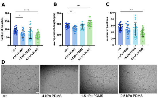 Fig.1 Alterations in endothelial tube architecture in response to changes in matrix stiffness.1
Customer Reviews
- "Enhanced Sensitivity in Anti-Angiogenic Screening. Using Creative Biolabs' endothelial tube formation & sprouting analysis service in our research has significantly improved our ability to distinguish between low-potency inhibitors. The automated junction counting was much more reliable than our previous manual methods." 12 days ago, S***n L.
- "Physiological Relevance in Wound Healing Models. Using Creative Biolabs' endothelial tube formation & sprouting analysis service in our research has significantly facilitated the development of our pro-angiogenic hydrogels. The 3D spheroid invasion data provided a clear correlation with our subsequent in vivo results." 25 days ago, M***ael K.
- "Streamlined High-Throughput Drug Testing. Using Creative Biolabs' endothelial tube formation & sprouting analysis service in our research has significantly facilitated our library screening. We processed over 50 compounds in record time with consistent results across multiple 96-well plates." 5 days ago, R***rt H.
FAQs
What is the main difference between your tube formation and sprouting assays?
The tube formation assay is a rapid 2D test focusing on how cells reorganize into networks, ideal for initial screening. The sprouting assay is a 3D model that measures how cells actually invade a matrix, which is more representative of physiological vessel growth. We can help you decide which is best for your specific drug target.
Can you perform these assays using primary cells from specific organs?
Yes, while HUVECs are the standard, we can perform the analysis using various primary endothelial cells, such as those from the lung, heart, or brain, to ensure organ-specific relevance for your project. Please reach out to discuss our current cell bank or shipping your own cells.
How do you ensure the reproducibility of the matrix used?
We use standardized lots of basement membrane extracts and verify the polymerization consistency for every batch. Our automated dispensing systems ensure that each well has an identical matrix thickness, which is crucial for comparable results.
Is it possible to study the interaction between endothelial cells and other cell types?
Absolutely. We offer co-culture services where we include pericytes, fibroblasts, or even tumor cells in the 3D matrix. This allows you to study how these "support" cells influence vessel stabilization or tumor-induced angiogenesis.
Related Services
Endothelial Cell Migration & Invasion Analysis Service
This service examines endothelial motility and invasive behavior in response to biochemical or matrix cues, helping characterize mechanisms involved in vascular remodeling and angiogenesis initiation.
Learn More →Microfluidic Vascular Modeling & Extravasation Simulation Service
This service utilizes microfluidic platforms to model perfusable vascular structures and cell extravasation behavior, enabling analysis of flow-dependent endothelial interactions and barrier function.
Learn More →Creative Biolabs provides an industry-leading endothelial tube formation & sprouting analysis service designed to deliver high-fidelity, quantitative data for your vascular research. From rapid 2D screening to complex 3D invasion models, we offer the technical expertise and automated platforms necessary to move your drug discovery pipeline forward with confidence.
ontact Our Team for More Information on Endothelial Tube Formation & Sprouting Analysis Service and to Discuss Your Project.
Reference
- Kretschmer, Maibritt et al. "Endothelial Sprout Formation Is Regulated by Substrate Stiffness and Notch Signaling." International journal of molecular sciences vol. 26,7 3155. 28 Mar. 2025. Distributed under Open Access license CC BY 4.0, without modification. https://doi.org/10.3390/ijms26073155
 Download our brochure
Loading case studies...
Online Inquiry