Primary or specialized endothelial cells are seeded into the channels and cultured under flow to develop a tight, mature monolayer.
Microfluidic Vascular Modeling & Extravasation Simulation Service
Are you currently facing poor clinical translation, high failure rates in oncology drug pipelines, or the limitations of static assays that fail to mimic hemodynamic forces? Our microfluidic vascular modeling & extravasation simulation service helps you obtain physiologically relevant data and develop highly specific therapeutic candidates through organ-on-a-chip technology, real-time perfusion modeling, and precise 3D matrix invasion analytics. We solve the challenge of accurately modeling complex vascular barriers in a high-throughput microfluidic environment.
Overview What We Can Offer Workflow Required Materials Highlights Customer Reviews FAQs Related Services
Overview
Microfluidic vascular modeling represents the frontier of biomimetic engineering, recreating the structural and mechanical complexity of human vasculature. Recent literature emphasizes that extravasation—the process of cells exiting the bloodstream—is highly dependent on shear stress and 3D matrix interactions. Traditional static models consistently underestimate the protective role of the endothelial barrier. Creative Biolabs integrates these advanced microfluidic principles to provide a validated, high-credibility platform for studying metastasis, inflammation, and drug delivery efficiency.
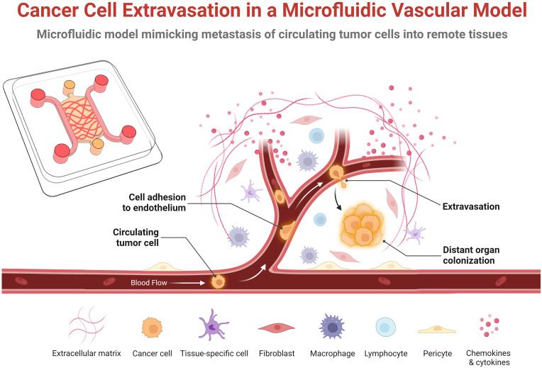 Fig.1 Overview of the main stages of cancer cell extravasation in a microfluidic vascular model.1
We utilize a multi-disciplinary strategy to simulate the vascular microenvironment with high fidelity. Our methodology focuses on the integration of micro-vessels within a tunable 3D extracellular matrix (ECM) to study the active crossing of the endothelial wall. We employ perfusion-based strategies that introduce controlled shear stress, mimicking physiological blood flow which is critical for endothelial cell alignment and barrier maturation. Furthermore, our spatial modeling includes the establishment of precise chemical gradients, such as chemokines or nutrients, to drive directional extravasation, allowing us to differentiate between passive paracellular leakage and active transcellular migration.
Empower Your Research with Creative Biolabs — Begin Your Expert Consultation Today
What We Can Offer
Real-Time Perfusion Modeling
Continuous flow control to simulate physiological hemodynamic forces and shear stress on endothelial monolayers.
3D Vascular Barrier Reconstruction
Assembly of perfusable micro-vessels within hydrogel scaffolds to replicate the basement membrane and interstitial space.
Extravasation Kinetic Tracking
High-resolution imaging to measure the rate and frequency of tumor cells or therapeutic agents exiting the vascular lumen.
High-Content Image Analytics
Automated quantification of junctional integrity, cell adhesion events, and 3D invasion depth within the matrix.
Pharmacological Barrier Validation
Testing of drug candidates to evaluate their ability to either protect the barrier or cross it effectively for targeted delivery.
Workflow
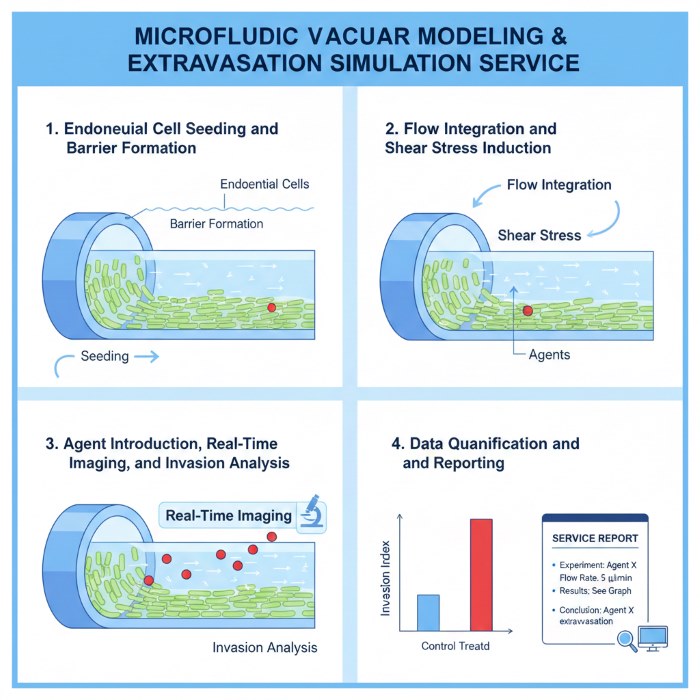
Endothelial Cell Seeding and Barrier Formation
Flow Integration and Shear Stress Induction
Automated pumps or pressure controllers are integrated to initiate perfusion, applying defined shear stress levels to the vessel wall.
Agent Introduction, Real-Time Imaging, and Invasion Analysis
Tumor cells, immune cells, or drug-loaded nanoparticles are introduced into the perfusate while live imaging captures their interaction with the endothelial barrier and subsequent translocation and accumulation within the surrounding 3D matrix.
Data Quantification and Reporting
Our team provides quantitative indices for extravasation rates, junctional protein expression, and barrier resistance.
Required Starting Materials
- Specific Cell Populations: Provision of your targeted tumor cells, immune cells, or specialized endothelial sources for barrier reconstruction.
- Therapeutic Candidate Profiles: Information on the molecular weight, solubility, and expected concentration ranges for the drug or nanoparticle candidates.
Highlights
Superior Biomimetic Hemodynamic Forces
Our advanced microfluidic systems recreate the absolute physiological shear stress found in human capillaries and arteries, ensuring that endothelial cells maintain their natural polarized phenotype.
Real-Time Kinetic Extravasation Resolution
By capturing the entire temporal process of cells exiting the vasculature, we identify the specific stages of adhesion and trans-endothelial migration that static measurements miss.

Advanced AI-Driven Barrier Quantification
We utilize proprietary image segmentation software to provide strictly objective metrics for junctional integrity and extravasation frequency, reducing human bias.
High-Throughput and Scale-Up Optimization
All microfluidic platforms are designed for parallelized operation, enabling the rapid screening of diverse compound libraries under flow.
Experience the Creative Biolabs Difference — Contact Us for a Tailored Quote
Customer Reviews
- "[High-Resolution Metastasis Insights] Using Creative Biolabs' microfluidic vascular modeling & extravasation simulation service in our research has significantly facilitated our study of breast cancer metastasis. Watching tumor cells extravasate in real-time provided invaluable mechanistic data." -2 months ago, S***n K.
- "[Unmatched Barrier Integrity Validation] Using Creative Biolabs' microfluidic vascular modeling & extravasation simulation service in our research has significantly facilitated our BBB-crossing nanoparticle project. The consistency of the endothelial junctions under flow was exactly what we needed." -10 days ago, A***a G.
FAQs
How does flow-induced shear stress affect extravasation results?
Shear stress is a primary mechanical cue that regulates endothelial gene expression and the formation of tight junctions. Without flow, the barrier is often "leakier" than in vivo, leading to false results.
Can you model specific tissue-tissue interfaces, like the Blood-Brain Barrier?
Yes, we specialize in co-culture chips that integrate endothelial cells with organ-specific cells (e.g., astrocytes for the BBB) across a porous membrane to replicate specific functions.
What is the advantage of microfluidics over traditional Transwell assays?
Transwells lack flow and shear stress. Microfluidics provides a dynamic, continuous-monitoring environment that much more closely mimics the human circulatory system.
Related Services
Angiogenesis-Tumor Microenvironment Interaction Modeling Service
This service models interactions between vascular structures and tumor-associated components, enabling assessment of how microenvironmental factors influence angiogenic behavior.
Learn More →Vascular Evaluation Profiling & Reporting Service
This service consolidates multi-level vascular data into structured profiles and interpretive reports, supporting clear comparison, decision-making, and downstream study planning.
Learn More →Our scientific team is ready to help you design a tailored study for your specific vascular barrier research. Whether you need an organ-specific chip or a high-throughput drug crossing screen, Creative Biolabs offers the support and expertise your project demands.
Contact Our Team for More Information on Microfluidic Vascular Modeling & Extravasation Simulation Service and to Discuss Your Project.
 Download our brochure
Loading case studies...
Online Inquiry