Creative Biolabs offers stable cell line development for monoclonal antibodies, CAR-T therapies, and drug screening. We optimize for high yields and long-term stability, ensuring correct glycosylation and protein folding.
Learn More →Reporter & Readout Validation Service for Cell Cycle Activity
Creative Biolabs provides a high-fidelity bridge between biological hypotheses and tangible kinetic data, ensuring fluorescent reporters accurately indicate phase boundaries. We resolve "temporal offsets" to pinpoint exact G1/S or S/G2 transitions, essential for characterizing checkpoint inhibitors or slow-cycling cancer stem cells. Our validation ensures synthetic reporters don't perturb endogenous cycles or metabolic health across models, including primary human intestinal epithelial cells (IECs).
Background What We Can Offer Workflow Publication Why Choose Us FAQs Customer Review Related Services Contact Us
Introduction to Advanced Cell Cycle Kinetics
The eukaryotic cell division cycle is a tightly orchestrated process essential for life and genome stability. Monitoring these transitions in living cells has traditionally been hampered by the limitations of static assays like flow cytometry. Modern research relies on fluorescent indicators like FUCCI to visualize these transitions; however, recent studies prove that original reporters can be variably offset from true S-phase boundaries. Creative Biolabs addresses this by implementing second-generation PIP-FUCCI and Ki67p-driven systems. Our validation services ensure that these reporters provide a precise demarcation of phase transitions, enabling the study of proliferation commitment and the maintenance of genome stability with single-cell resolution.
Comprehensive Cell Cycle Tracking Capabilities
Creative Biolabs offers a diverse portfolio of validated reporter systems tailored to specific research objectives. Our "What We Offer" section is designed to match the right tool to your biological question:
Second-Generation PIP-FUCCI Systems
Utilizing the PIP-degron motif, these reporters are directly coupled to DNA replication machinery. This is our most recommended tool for drug discovery projects requiring the highest temporal precision at the G1/S boundary.
Ki67p-FUCCI for Quiescence Mapping
We specialize in promoter-driven systems that utilize the Ki67 proximal promoter. This allows for the clear differentiation between the G0 (quiescent) and G1 (proliferative) states, which is essential for research into cellular dormancy and regenerative medicine.
CDK-Activity Translocation Reporters (KTR)
For projects focused on signaling hubs, we validate KTRs that convert cyclin-dependent kinase activity into a nucleocytoplasmic translocation readout, providing a continuous measure of kinase dynamics throughout the cycle.
PCNA-Variance Ground-Truth Analysis
We offer co-expression validation using mTurquoise2-PCNA. By calculating the spatially localized variance of PCNA puncta, we provide a definitive "ground truth" calibration for any other cell cycle reporter in your pipeline.
Explore the Full Spectrum of Our Validated Technologies — Consult with a Cell Engineering Specialist
Service Workflow and Technical Execution
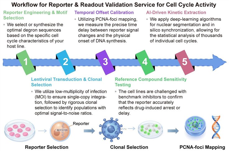
Our validation process is structured to provide complete transparency and scientific rigor at every stage:
Publication
This review provides a concise guide to genetically encoded fluorescent reporters for live-cell imaging of the cell cycle. It compares the molecular basis, strengths, and limitations of major systems, including FUCCI, kinase translocation reporters (KTRs), and PCNA-based replication foci reporters. The article highlights how each tool captures distinct dynamics and discusses strategies for combining multiple reporters to gain a more comprehensive, accurate understanding of cell cycle progression and regulation in single living cells.
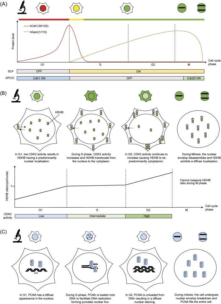 Fig.1 The molecular basis and fluorescence imaging output of cell cycle reporters.1
Why Choose Us?
Creative Biolabs distinguishes itself by solving the "growth-cycle paradox"—the difficulty of determining whether a change in reporter signal is due to a specific cell cycle block or a general change in cellular growth rate. Unlike standardized kits, our service is highly customized. We utilize advanced PIP-FUCCI and Ki67p architectures that offer resolution far beyond original FUCCI systems. Our expertise in primary human cells and organoid-compatible imaging ensures that your validation is performed in a physiologically relevant context. Furthermore, our platform is supported by Published Data demonstrating our ability to decouple fluorescent readouts from stress-induced artifacts. Whether you are working with metazoan cancer models or the fission yeast S. pombe, our cross-species expertise ensures a robust and reliable validation.
Partner with Industry Leaders to Elevate Your Research — Contact Us for a Custom Quote
FAQs
How do you ensure the reporter doesn't interfere with normal cell growth?
We perform rigorous growth-curve analysis. We guarantee that the doubling time of our validated lines remains within ±5% of the parental line, ensuring that the reporter is biologically neutral.
What makes PIP-FUCCI superior to the original FUCCI system?
Original FUCCI often shows a signal lag, making it hard to distinguish early S-phase from G1. PIP-FUCCI is directly coupled to DNA replication via the PIP-degron, offering near-instantaneous degradation at the start of S-phase.
Can this service be applied to primary human cells or organoids?
Absolutely. We have developed specialized protocols for viral transduction and imaging in primary human cells, including the use of collagen-matrix platforms to maintain physiological proliferative capacity.
Customer Review
-
Unprecedented Accuracy in G1/S Mapping
Using Creative Biolabs' PIP-FUCCI validation in our research has significantly facilitated our understanding of DNA damage-induced G1 arrest. The PCNA-variance correlation corrected a 2-hour offset we had in our previous unvalidated model. - Dr. Al***n M -
Seamless Primary Cell Integration
Using Creative Biolabs' reporter validation in our research has significantly improved the stability of our human intestinal organoid models. Their matrix-optimized imaging was crucial for our long-term time-lapse studies. - Prof. S***ra H
Related Services
Cell Line Development Service
Phage Display based Antibody Discovery Service
Creative Biolabs constructs massive antibody libraries displayed on filamentous phages to screen for high-affinity drugs and vaccines. Our high-efficiency biopanning enables rapid epitope mapping and therapeutic discovery.
Learn More →How to Contact Creative Biolabs
Creative Biolabs provides an end-to-end reporter and readout validation service for cell cycle activity, transforming live-cell imaging into a high-fidelity quantitative tool. Our expertise in PIP-FUCCI, Ki67p-quiescence mapping, and AI-driven PCNA-variance analysis ensures that your drug discovery pipeline is built on accurate, biological ground truth.
For more information or to discuss the specific requirements of your cell cycle project, please reach out to our scientific team. We are ready to provide a detailed project proposal tailored to your cell model.
Reference
- Wang, Jinyu, et al. "A concise guide to fluorescent cell cycle reporters for live-cell imaging." Frontiers in cell and developmental biology 13 (2025): 1702230. Distributed under Open Access license CC BY 4.0, without modification. https://doi.org/10.3389/fcell.2025.1702230
 Download our brochure
Loading case studies...
Online Inquiry