Creative Biolabs provides orthogonal validation to de-risk WIP1 (PPM1D) modulators. We eliminate chemical interference and substrate bias, providing mechanistic clarity on gatekeeper functions to accelerate candidate transition.
Learn More →Spatial Cell Cycle Activity Mapping Service
Creative Biolabs provides a high-resolution spatial census to identify kinetic trajectories within complex tissues. By mapping the exact coordinates of cell cycle phases, we reveal niche-dependent regulatory mechanisms invisible to bulk analysis. This platform identifies proliferative hotspots and quiescent zones shielding cells from therapeutics. Furthermore, we characterize the proliferation-differentiation transition, such as mapping B-cell polarization within germinal centers. Our spatial datasets provide definitive evidence for target engagement and high-impact publications.
Background What We Can Offer Workflow Publication Why Choose Us FAQs Customer Review Related Services Contact Us
Spatial Cell Cycle Activity Mapping: Accelerate Your Drug Discovery Process!
Do you find it difficult to validate cell cycle transitions in complex tissues, or are you losing vital spatial context through traditional sequencing? Relying on dissociated cells often masks the relationship between a cell's proliferative state and its immediate microenvironment, resulting in fragmented efficacy data. Creative Biolabs offers spatial cell cycle activity mapping to bridge this gap. By combining subcellular transcriptomics with high-plex protein detection, we visualize how localized signaling gradients and metabolic stress trigger the switch between division and dormancy. Our platform uncovers resistant populations and niche-dependent dynamics, providing the context-aware insights necessary to optimize your therapeutic strategies.
Comprehensive Capabilities
To ensure the highest degree of functional validation, Creative Biolabs offers a modular and highly customizable service suite. Our offerings are designed to translate complex spatial data into actionable biological insights for your research pipeline.
Subcellular Phase Resolution
Utilizing diffraction-limited and super-resolution imaging, we map mRNA transcripts with <1 µm precision. This allows for the observation of nuclear versus cytoplasmic localization of cell cycle regulators, providing clues into the activation state of specific checkpoints.
Customizable "Cell Cycle Sensor" Panels
We offer a pre-validated library of over 100 cell cycle-related probes across human, mouse, and non-human primate (NHP) species. Furthermore, we can design and synthesize custom probes for your specific therapeutic targets or lineage-specific markers to see how your drug directly influences the cycle in situ.
Localized Hypoxia and Metabolic Profiling
We can co-map cell cycle activity with markers of metabolic stress and oxygen tension (e.g., HIF1A, LDHA). This enables the identification of "dormant zones" where metabolic restrictions may be driving cell cycle exit and therapeutic persistence.
Spatial Pseudotime & Kinetic Trajectory Modeling
Rather than providing a static "snapshot," our bioinformatics team models the continuous flow of cells through the cycle. By analyzing the transcriptomic gradients across tissue coordinates, we can estimate the relative time cells spend in specific phases within different microenvironments.
Neighborhood Enrichment Analysis
We provide a detailed statistical breakdown of the "cell-to-cell" interactions driving proliferation. This includes quantifying the proximity of cycling cells to regulatory immune cells or specific stromal components that may be providing pro-proliferative signals.
Inquire About Our Custom Panel Design Options - Get Started with a Technical Assessment
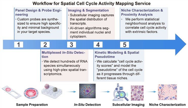
Detailed Workflow and Technical Implementation
To initiate the service, Creative Biolabs coordinates closely with your team to ensure that experimental parameters are optimized for the specific biology of your target tissue.
Publication
This publication introduces CMAP (cellular mapping of attributes with position), a computational method that integrates single-cell and spatial transcriptomics data through a three-level mapping strategy. It assigns precise spatial coordinates to individual cells, overcoming the resolution limitations of current spatial technologies. CMAP demonstrates robust performance across diverse datasets and platforms, enabling high-resolution analysis of spatial heterogeneity, such as organ-specific endothelial cell variation and complex tumor immune microenvironments.
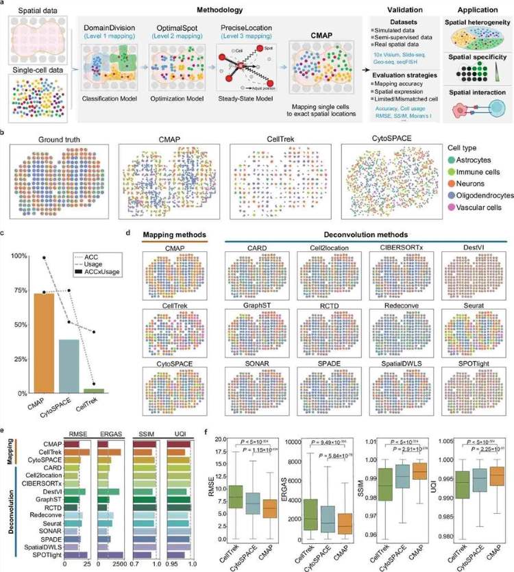 Fig.1 Framework construction and application assessment of the CMAP method for cellular attribute-position mapping.1
Why Choose Us?
Partnering with Creative Biolabs provides a continuous model of cell cycle activity, transcending the limitations of traditional immunohistochemistry (IHC) snapshots. Our platform integrates transcriptomic "intent" with proteomic "action," delivering multi-layered biological evidence. We offer superior sensitivity via specialized signal amplification, crucial for identifying elusive G1/M transitions. Furthermore, our specialized quiescence detection distinguishes G0 populations by analyzing S-phase downregulation alongside niche-specific dormancy markers. Grounded in peer-reviewed spatial frameworks, our methodologies ensure evidence-based success and high-fidelity functional validation for your most complex biological questions.
Experience the Creative Biolabs Advantage - Get a Personalized Quote for Your Project Today
FAQs
How does this service compare to traditional Ki-67 IHC?
While Ki-67 provides a binary "proliferating vs. non-proliferating" view, our service utilizes a multi-gene panel to resolve specific phases (G1, S, G2, M) and uses spatial pseudotime to model the speed of the cycle, providing much deeper functional context for drug response analysis.
Is it possible to integrate my specific therapeutic target into the panel?
Absolutely. We can customize panels to include up to 50+ additional markers, allowing you to correlate cell cycle activity directly with the spatial expression of your drug target or specific resistance markers.
How do you validate the transcriptomic cell cycle predictions?
We provide integrated multi-omics validation by co-detecting phase-specific proteins alongside the mRNA sensors to ensure the predictions are biologically grounded.
Customer Review
-
Unprecedented Niche Insights
Using Creative Biolabs' spatial cell cycle mapping in our tumor microenvironment research has significantly facilitated our understanding of G0-entry triggers. The spatial pseudotime analysis provided a level of kinetic detail we couldn't achieve with flow cytometry. - Dr. Al***rt S -
Accurate Dormancy Detection
We were struggling to find where our cells were exiting the cell cycle post-treatment. Using this service, we mapped the exact spatial coordinates of quiescence, which led to a new hypothesis regarding stromal interference. - Dr. Ke***n W
Related Services
Orthogonal Molecular Assay
IHC-based Tumor Profiling
Creative Biolabs provides high-quality IHC services, offering expert paraffin/OCT embedding, sectioning, and staining. Our pathologists deliver rapid, precise biomarker analysis and interpretation for comprehensive final reports.
Learn More →How to Contact Creative Biolabs
Creative Biolabs is dedicated to providing the most advanced spatial biology tools to support your drug discovery and functional validation needs. Our team of specialists is ready to help you navigate the complexities of tissue heterogeneity and cellular dynamics.
Contact our specialists today to discuss your project requirements and receive a customized technical proposal.
Reference
- Ke, Jincan, et al. "High-resolution mapping of single cells in spatial context." Nature Communications 16.1 (2025): 6533. Distributed under Open Access license CC BY 4.0, without modification. https://doi.org/10.1038/s41467-025-61667-4
 Download our brochure
Loading case studies...
Online Inquiry