
Creative Biolabs is offering the most comprehensive services for antibody development projects. With strict regulation and effective execution, we are dedicated to providing the most valuable solutions to complete your projects.
HighlightsCreative Biolabs is offering the most comprehensive services for antibody development projects. With strict regulation and effective execution, we are dedicated to providing the most valuable solutions to complete your projects.
HighlightsCreative Biolabs has established a broad range of platforms for developing novel antibodies or equivalents. These cutting-edge technologies enable our scientists to meet your demands from different aspects and tailor the most appropriate solution that contributes to the success of your projects.
HighlightsWith deep understanding in antibody-related realms and extensive project experience, Creative Biolabs offers a variety of references to help you learn more about our capacities and achievements, including infographic, flyer, case study, peer-reviewed publications, and all kinds of knowledge that can assist your projects. You are also welcome to contact us directly for more specific solutions.
HighlightsGet a real taste of Creative Biolabs, one of the most professional custom service providers in the world. We are committed to providing highly customized comprehensive solutions with the best quality to advance your projects.
HighlightsIn the rapidly evolving landscape of precision medicine and targeted drug delivery, the vascular endothelium stands as the most critical biological interface. As the primary barrier and gateway between the systemic circulation and the parenchyma of every organ, the endothelium expresses a highly heterogeneous array of molecular "ZIP codes" that define tissue identity and disease states. Creative Biolabs, a global leader in phage display technology with over two decades of specialized expertise, offers the most comprehensive Vascular Endothelium Targeting In Vivo Phage Library Screening Service in the industry. Our platform integrates high-diversity combinatorial libraries, advanced animal models, and Next-Generation Sequencing (NGS) to map the vascular landscape with unprecedented resolution.
The vascular endothelium is not merely a passive lining of blood vessels; it is a dynamic, multi-functional organ. Its molecular composition varies significantly across different anatomical sites, influenced by local hemodynamics, oxygen tension, and tissue-specific signaling. This molecular diversity provides a unique opportunity for In Vivo Phage Display for Vascular Endothelium, allowing for the discovery of ligands that can bypass systemic degradation and home specifically to a target organ or pathological lesion.
The concept of vascular "addressing" suggests that endothelial cells in the brain, lung, heart, or tumors express distinct surface markers (receptors, integrins, or peptidases). By utilizing In Vivo Phage Library Screening, we can identify the specific keys (peptides or scFv/Fab fragments) that fit these molecular locks.
Beyond physiological diversity, disease states such as cancer, inflammation, and atherosclerosis induce the expression of "neo-addresses." For instance, tumor-associated vessels often overexpress aminopeptidase N (CD13) or αvβ3 integrins. Our screening service is designed to isolate ligands that specifically recognize these upregulated markers, providing a foundation for targeted chemotherapy, gene therapy, and molecular imaging.
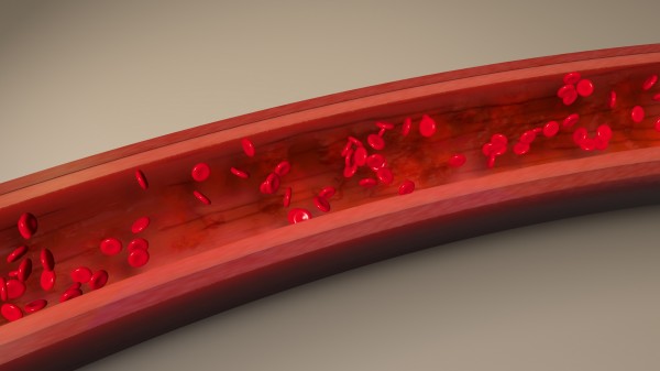 Fig. 1 Blood vessel endothelium layer damage.
At Creative Biolabs, we have transitioned beyond traditional biopanning. Our service suite incorporates the latest advancements in biotechnology to ensure the highest specificity and translatability of identified clones.
The success of In Vivo Phage Library Screening depends on the initial library's quality and diversity. We offer:
To ensure clinical relevance, we perform screenings in a variety of validated models:
Traditional "Sanger-only" screening often misses rare but high-affinity clones. Our platform utilizes:
Our workflow for Vascular Endothelium Targeting In Vivo Phage Library Screening is meticulously designed to maximize the recovery of organ-specific binders while minimizing non-specific background.

Before injection, the library is carefully characterized. In some cases, a "negative selection" or "subtractive panning" step is performed in vitro against non-target cells (e.g., hepatocytes or common fibroblasts) to remove ubiquitous binders.
The phage library is injected intravenously (usually via the tail vein in rodents). The phages circulate through the entire vascular tree, allowing them to interact with the luminal surface of the endothelium under native physiological flow conditions.
We optimize the circulation time (ranging from minutes to hours) based on the target. This allows high-affinity phages to bind their respective endothelial receptors. Our platform also includes "internalization-focused" protocols for ligands that must enter the endothelial cell or cross the Blood-Brain Barrier (BBB).
This is a critical step where Creative Biolabs excels. To remove non-specifically bound or circulating phages, the animal is subjected to extensive systemic perfusion with saline or buffered solutions. This ensures that only the phages tightly bound to the vascular wall are recovered.
Target organs (and control organs) are harvested. We utilize various elution techniques:
The recovered phages are amplified and subjected to subsequent rounds of in vivo screening (typically 3-4 rounds). NGS is performed at each round to generate a "fitness landscape" of the library enrichment.
Candidate clones are validated through:
Creative Biolabs has successfully mapped various vascular "ZIP codes." Our In Vivo Phage Display expertise extends to the following high-demand areas:
Identifying peptides that can trigger receptor-mediated transcytosis across the BBB is the "holy grail" of CNS drug delivery. We focus on receptors like Transferrin (TfR) and LRP1, but also discover novel, uncharacterized pathways.
The lung's massive surface area makes it an ideal target for treating respiratory distress, pulmonary hypertension, and metastatic cancer. We have identified motifs specific to the lung endothelium that avoid liver and spleen sequestration.
Cancerous blood vessels are structurally and molecularly distinct. We screen for ligands targeting:
Table 1. Comparison of Targeting Efficiency across Organ
| Target Organ | Common Surface Marker | Enrichment Ratio (Typical) | Lead Motif Example |
| Brain | LRP1, TfR, GLUT1 | 10-50x | THR, Angiopep-2 |
| Lung | Aminopeptidase P | 20-100x | GFE |
| Tumor | CD13, αvβ3 | 50-500x | RGD, NGR |
| Kidney | Megalin | 5-20x | LTS |
As the industry's premier provider of In Vivo Phage Library Screening Services, Creative Biolabs provides unparalleled technical depth:
To further your research, consider our integrated In Vivo Phage Library Screening Services modules:
Learn more about other Phage Display Library Screening Services:
Creative Biolabs is dedicated to pushing the boundaries of what is possible in vascular targeting. Our expert team is ready to discuss how our Vascular Endothelium Targeting In Vivo Phage Library Screening Service can accelerate your drug discovery program.
References
All listed services and products are For Research Use Only. Do Not use in any diagnostic or therapeutic applications.








