
Creative Biolabs is offering the most comprehensive services for antibody development projects. With strict regulation and effective execution, we are dedicated to providing the most valuable solutions to complete your projects.
HighlightsCreative Biolabs is offering the most comprehensive services for antibody development projects. With strict regulation and effective execution, we are dedicated to providing the most valuable solutions to complete your projects.
HighlightsCreative Biolabs has established a broad range of platforms for developing novel antibodies or equivalents. These cutting-edge technologies enable our scientists to meet your demands from different aspects and tailor the most appropriate solution that contributes to the success of your projects.
HighlightsWith deep understanding in antibody-related realms and extensive project experience, Creative Biolabs offers a variety of references to help you learn more about our capacities and achievements, including infographic, flyer, case study, peer-reviewed publications, and all kinds of knowledge that can assist your projects. You are also welcome to contact us directly for more specific solutions.
HighlightsGet a real taste of Creative Biolabs, one of the most professional custom service providers in the world. We are committed to providing highly customized comprehensive solutions with the best quality to advance your projects.
HighlightsIn the contemporary landscape of biomedical research, the zebrafish has emerged as an indispensable vertebrate model system for studying developmental biology, toxicology, and human disease pathogenesis. To empower researchers with high-specificity tools, Creative Biolabs provides a comprehensive, end-to-end Zebrafish Monoclonal Antibody Discovery service. Leveraging our world-leading phage display technology and decades of expertise in immune antibody library engineering, we deliver high-affinity Zebrafish Monoclonal Antibodies tailored to your specific research and diagnostic (non-clinical) needs.
Zebrafish offer a unique immunological repertoire. Unlike mammalian models, teleost fish utilize three distinct immunoglobulin (Ig) classes: IgM, IgD, and the fish-specific IgZ (analogous to mammalian IgA in mucosal immunity). Utilizing Zebrafish Monoclonal Antibody Discovery allows for the identification of binders that recognize epitopes often conserved across species or those that are non-immunogenic in traditional rodent models.
At Creative Biolabs, we specialize in the construction and screening of immune antibody library systems derived from zebrafish. By utilizing phage display-based Zebrafish Monoclonal Antibody discovery, we bypass the limitations of traditional hybridoma technology—such as low fusion efficiency and the instability of fish-mouse heterohybridomas—to provide a robust, recombinant solution for antibody production.
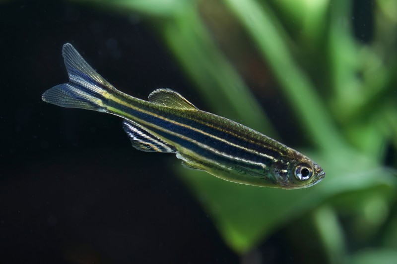
The cornerstone of our service is the integration of phage display with a sophisticated immune antibody library for Zebrafish Monoclonal Antibody discovery. Our platform is designed to capture the full diversity of the zebrafish immune response following antigen challenge.
Phage display involves the fusion of antibody fragments (typically scFv or Fab) to the coat proteins (pIII or pVIII) of filamentous bacteriophages. This creates a physical link between the phenotype (antibody binding) and the genotype (the DNA sequence). For Zebrafish Monoclonal Antibody projects, we clone the V-gene repertoire from zebrafish B-lymphocytes into phagemid vectors, ensuring that the resulting immune antibody library accurately reflects the animal's natural immune response.
Our Zebrafish Monoclonal Antibody Discovery process is a meticulously orchestrated pipeline, ensuring the highest success rate for even the most challenging antigens.
Successful Zebrafish Monoclonal Antibody generation begins with optimal immunization. We utilize high-purity proteins, peptides, or even whole cells as immunogens. Our proprietary adjuvant formulations are optimized for teleost fish to maximize the titers of IgM and IgZ-secreting B cells.
Following a confirmed immune response (monitored via ELISA), we harvest the spleen and kidney (the primary hematopoietic organs in zebrafish). Total RNA is extracted to provide a template for cDNA synthesis, targeting the immunoglobulin heavy and light chain constant regions.
Using a comprehensive set of degenerate primers specifically designed for zebrafish V-gene families (VH, VL, Vλ), we amplify the variable regions.
Our construction and screening of immune antibody library protocols ensure a library size of at least 108 to 109 independent clones, capturing the maximum depth of the immune repertoire.
We employ stringent biopanning strategies to isolate high-affinity binders from the immune antibody library for Zebrafish Monoclonal Antibody.
Selected clones are expressed as soluble proteins and subjected to rigorous validation, including:
Creative Biolabs stands at the forefront of Zebrafish Monoclonal Antibody Discovery. By combining the power of phage display with our deep expertise in immune antibody library technology, we provide the global scientific community with high-performance tools to unlock the secrets of zebrafish biology. Our commitment to quality and innovation ensures that your research is supported by the finest Zebrafish Monoclonal Antibodies available in the industry.
While our phage display immune antibody library platform is a powerful engine, we offer a comprehensive suite of discovery technologies to ensure we always use the right tool for the job. Our Monoclonal Antibody Generation from Various Immunized Antibody Libraries Services:
Contact us today to discuss your specific requirements for Zebrafish Monoclonal Antibody Discovery by phage display and let our experts design a customized strategy for your success.
Reference
All listed services and products are For Research Use Only. Do Not use in any diagnostic or therapeutic applications.








