
Creative Biolabs is offering the most comprehensive services for antibody development projects. With strict regulation and effective execution, we are dedicated to providing the most valuable solutions to complete your projects.
HighlightsCreative Biolabs is offering the most comprehensive services for antibody development projects. With strict regulation and effective execution, we are dedicated to providing the most valuable solutions to complete your projects.
HighlightsCreative Biolabs has established a broad range of platforms for developing novel antibodies or equivalents. These cutting-edge technologies enable our scientists to meet your demands from different aspects and tailor the most appropriate solution that contributes to the success of your projects.
HighlightsWith deep understanding in antibody-related realms and extensive project experience, Creative Biolabs offers a variety of references to help you learn more about our capacities and achievements, including infographic, flyer, case study, peer-reviewed publications, and all kinds of knowledge that can assist your projects. You are also welcome to contact us directly for more specific solutions.
HighlightsGet a real taste of Creative Biolabs, one of the most professional custom service providers in the world. We are committed to providing highly customized comprehensive solutions with the best quality to advance your projects.
HighlightsIn the rapidly evolving landscape of biotherapeutics and proteomics, the demand for sophisticated phage display libraries has never been higher. While the M13 filamentous phage system remains a staple for small peptide and scFv display, it often falters when faced with large, complex, or multi-subunit proteins.At Creative Biolabs, we bridge this gap through our world-class T4 Phage Library Construction Services. By leveraging the unique structural biology of the T4 Phage, we provide a robust platform capable of displaying high-molecular-weight proteins and enzymes that are traditionally "undisplayable" in other systems. With over 20 years of expertise, our team delivers high-diversity libraries (>1010) tailored for the most challenging research objectives.
The T4 Phage (Escherichia virus T4) is a large, complex virus that infects E. coli. Unlike the slender M13 phage, T4 possesses a massive prolate head (capsid). This structural volume, combined with its unique assembly pathway, offers unprecedented advantages for T4 Phage Library construction.
The Role of HOC and SOC Proteins
The T4 capsid is decorated with two non-essential "accessory" proteins:
By fusing your target of interest to the N- or C-terminus of HOC or SOC, Creative Biolabs can display hundreds of copies of a protein on a single phage particle. This high-density display is critical for increasing avidity during the screening of phage display libraries.
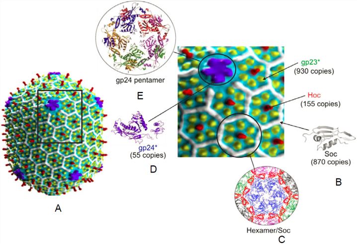 Fig 1. Structure of the bacteriophage T4 head.1
Our T4 Phage Library Construction platform is specifically engineered to overcome the "secretion bottleneck" of traditional systems.
Cytoplasmic Assembly (No Secretion Required)
Most phage systems (like M13) require the displayed protein to be secreted across the inner membrane into the periplasm. This often leads to misfolding or toxicity. T4 Phage assembles entirely within the host cytoplasm. The phage particles are released via cell lysis, meaning:
Massive Payload Capacity
The T4 system can accommodate massive DNA inserts (up to 170 kb). This allows for the construction of libraries containing:
Enhanced Stability
T4 phage particles are remarkably resilient to environmental stressors, including pH fluctuations and detergents. This allows for "stringent biopanning," ensuring that only the highest-affinity binders are selected from your T4 Phage Library.
To help you select the optimal system, the following table compares T4 with other common phage display platforms.
Table 1. Comparison of Phage Display Systems
| Feature | M13 Phage | T7 Phage | T4 Phage |
| Capsid Symmetry | Filamentous | Icosahedral | Prolate Icosahedral |
| Assembly Site | Periplasm (Secretion) | Cytoplasm (Lysis) | Cytoplasm (Lysis) |
| Genome Type | ssDNA | dsDNA | dsDNA (169 kb) |
| Display Site | pIII, pVIII | Capsid 10B | HOC, SOC |
| Max Insert Size | Small (<50 kDa) | Medium | Large (>100 kDa) |
| Copy Number | 3–5 (pIII) | 0.1–415 | 155 (HOC) / 810 (SOC) |
| Secretion Stress | High | Low | None |
Our process is rigorous, transparent, and fully customizable.
As a leader in the industry, Creative Biolabs integrates the latest biotechnologies into our T4 Phage Library Construction workflow.
AI-Driven Library Design
We utilize machine learning algorithms to predict the optimal fusion sites on HOC/SOC and to design "smart" libraries that minimize sequence bias while maximizing structural stability.
Assisted Engineering
Our team employs genome editing to precisely modify the T4 genome, allowing for the creation of "empty" HOC/SOC platforms or the introduction of specific metabolic markers for easier tracking and selection.
In Vitro Packaging (IVP) Systems
To bypass the limitations of bacterial transformation efficiency, we offer in vitro packaging. This allows us to achieve library diversities exceeding 1011 by directly packaging recombinant DNA into pre-formed T4 phage heads in a cell-free environment.
With 20+ years in the industry, Creative Biolabs is more than just a service provider; we are your collaborative partner in science.
To further your research, consider our integrated phage display library construction service modules:
Learn more about other T4 phage library construction services:
Creative Biolabs' T4 Phage Library Construction Services provide the power, capacity, and precision you need to discover the next generation of therapeutics. Contact our experts today to discuss your project requirements and receive a detailed quote.
Reference
All listed services and products are For Research Use Only. Do Not use in any diagnostic or therapeutic applications.








