Irrespective of the methods utilized for diagnostic imaging, adequate signal intensity in a specific region must be achieved. It is used to differentiate a specific region from its surroundings. Liposomes are efficient molecular imaging probe carriers. Liposome-based imaging probes can promote the accumulation of probes in particular regions during disease diagnosis, enhancing the signal differential between sick and normal tissues. Utilizing liposome-based Imaging probes, we can meet your diagnostic needs for brain, lung, liver, spleen, cardiovascular system, tumors, and more.
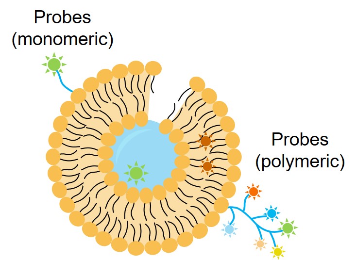 Fig 1. Liposome binding to imaging probes
Liposome-based CT Probes
Computed tomography (CT) imaging offers a great spatial resolution and limitless penetration depth. But it has a poor sensitivity, which necessitates adequate imaging agent administration. Loading imaging probes with liposomes could prolong blood circulation time. For imaging specific tissues, we can provide you with liposome-based CT probes loaded with iodine, gold nanoparticles, and so on.
Liposome-based MRI Probes
Magnetic resonance imaging (MRI) provides excellent spatial resolution and multi-sequence imaging for simultaneous acquisition of anatomical and physiological information, but it is limited in sensitivity. We can co-load drugs with imaging probes (e.g., gadolinium and iron oxide) in liposomes to constitute lipid-based MRI probes. This strategy can improve dtug delivery with image guidance and extend the blood circulation period of the imaging probe, hence improving the image signal.
Liposome-based USI Probes
Ultrasound imaging (USI) may image soft tissue by comparing the sound signals returned by different tissues. It has the advantages of real-time operation, portability, inexpensiveness, high spatial resolution, and no dangerous ionizing radiation. By encapsulating bubbles within liposomes, our liposome-based USI probes can increase echogenicity of blood.
Liposome-based FLI Probes
Fluorescence imaging (FLI) is a real-time imaging technique that uses fluorescent dyes such as ICG, IR800-CW, IR780, rhodamine, and porphyrin to label target cells and tissues. Fluorescent dyes encapsulated in liposomes have higher cellular uptake and longer blood circulation time than free fluorescent dyes. We can provide you with liposome-based FLI probes for synergistic photothermal/chemotherapy by triggering drug release under light while fulfilling image-guided photothermal and/or photodynamic therapy.
Liposome-based PAI Probes
Photoacoustic imaging (PAI) is a biomedical modality based on the photoacoustic effect, where light is absorbed in a sample to form sound waves. We can provide you with liposome-based PAI probes. The probes encapsulate fluorescent dyes with PA signaling such as Bchl-lipid, ICG, and others. As a result, PAI probes can provide photothermal effects to guide photothermal therapy.
Liposome-based multimodal imaging probes
Each imaging method has benefits and drawbacks. We may develop liposome-based multimodal imaging probes by combining multiple imaging modalities (e.g., FLI/MRI, FLI/PET, FLI/CT/MRI, FLI/PAI/MRI). This will help you learn more about your target cells/tissues.
Creative Biolabs is committed to providing cutting-edge solutions for molecular diagnosis using a wide range of imaging modalities including CT, MRI, USI, PET, multimodal imaging, etc. Several innovative liposome-based products (e.g., biomimetic membrane-based targeting probes, liposome-based target activatable probes) have been developed by our expert group to enhance the sensitivity and specificity of these techniques. We can help you achieve unprecedented accuracy and precision in the diagnosis imaging process. Please contact us for the most appropriate solution if you have any questions about imaging or diagnosis.
 For Research Use Only. Not For Clinical Use
Applications
Online Inquiry
Creative Biolabs is a world-leading liposome provider with extensive experience in liposome preparation, custom formulation development, and characterization.

© 2026 Creative Biolabs. All rights reserved.