As a leading source provider in the field of biological research and drug discovery, Creative Biolabs is fully competent and dedicated to providing a range of phospholipid-based pharmaceutical formulations and related services. As a service provider, our goal is to provide the highest level of service, confidentiality and customer support.
The anionic phospholipid phosphatidylglycerol (PG) is an important phospholipid found by Benson and Maruo from Scenedesmus cells in 1958. Since this discovery, PG has been found in many organisms, and it is now considered to be a ubiquitous phospholipid that is present in almost all organisms.
In mammals, PG is a minor phospholipid component of many intracellular membranes, accounting for less than 1% of total phospholipids, and it's predominantly located in mitochondria and microsomal membranes. In the lung, PG is one of the major components of lung surfactants, mainly in the lamellar membrane. In cyanobacteria and chloroplasts of higher plants, most of the PG is present in the thylakoid membrane, in which oxygenic photosynthesis takes place. PG typically represents a minor component next to other phospholipids, but in the thylakoid and inner envelope, PG is the only phospholipid present and it is present at low levels. In both prokaryotes and eukaryotes, PG is synthesized by cytosine 5’-diphosphate (CDP)-diacylglycerol and glycerol-3-phosphate, involving the action of a membrane-bound PG-phosphate synthase and PG-phosphate phosphatase. In plants, PG biosynthesis occurs in the inner envelope membranes of plastids, in the inner membranes of mitochondria and in the endoplasmic reticulum.
The phosphate group in PG is negatively charged at neutral pH, so PG is classified as an anionic phospholipid. The PG structure is shown in Figure 1. In cyanobacteria and in the chloroplasts of higher plants that perform oxygen-containing photosynthesis, most of the PG is found in the thylakoid membrane, which is the site of photosynthetic light reactions and electron transport. Therefore, PG may play an important role in the main process of photosynthesis in thylakoid membranes. In non-photosynthetic prokaryotes, such as Escherichia coli, PG is an important but not essential participant in cellular metabolism. In non-photosynthetic eukaryotes, PG is synthesized only in mitochondria, where it is involved in the biosynthesis of cardiolipin, which is mainly located in the inner membrane of mitochondria.
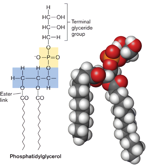 Fig 1 Structural model of phosphatidylglycerol.
Our Service for Phosphatidylglycerol
Creative Biolabs is fully committed to providing PG related best-customized formulations and services to customers around the world. Our services have the following advantages:
With top-notch technologies and a professional team, Creative Biolabs provides a variety of services based on drug delivery, pharmaceutical preparations and more to customers around the world. Our dedicated technical support team ensures that we provide the best service available for every customer, and we make sure that you can achieve your goals here.
 For Research Use Only. Not For Clinical Use
Supports
Online Inquiry
Creative Biolabs is a world-leading liposome provider with extensive experience in liposome preparation, custom formulation development, and characterization.

© 2026 Creative Biolabs. All rights reserved.