
Creative Biolabs is offering the most comprehensive services for antibody development projects. With strict regulation and effective execution, we are dedicated to providing the most valuable solutions to complete your projects.
HighlightsCreative Biolabs is offering the most comprehensive services for antibody development projects. With strict regulation and effective execution, we are dedicated to providing the most valuable solutions to complete your projects.
HighlightsCreative Biolabs has established a broad range of platforms for developing novel antibodies or equivalents. These cutting-edge technologies enable our scientists to meet your demands from different aspects and tailor the most appropriate solution that contributes to the success of your projects.
HighlightsWith deep understanding in antibody-related realms and extensive project experience, Creative Biolabs offers a variety of references to help you learn more about our capacities and achievements, including infographic, flyer, case study, peer-reviewed publications, and all kinds of knowledge that can assist your projects. You are also welcome to contact us directly for more specific solutions.
HighlightsGet a real taste of Creative Biolabs, one of the most professional custom service providers in the world. We are committed to providing highly customized comprehensive solutions with the best quality to advance your projects.
HighlightsThe development of in vitro display technologies transformed molecular biology through the ability to select high-affinity peptides and proteins that do not depend on living cells. By connecting genetic information to physical traits researchers can quickly develop biomolecules with specific desired features.
The most prominent in vitro display systems include:
These platforms underpin modern protein engineering, antibody discovery, and therapeutic design.
As protein therapeutics continue to dominate the biopharmaceutical landscape, display technologies remain at the forefront of innovation. Their capacity to identify rare, high-affinity binders from vast libraries makes them essential for:
Phage display links peptides or proteins to their encoding DNA by fusing them to a coat protein (commonly pIII or pVIII) of filamentous bacteriophages like M13. The process involves:
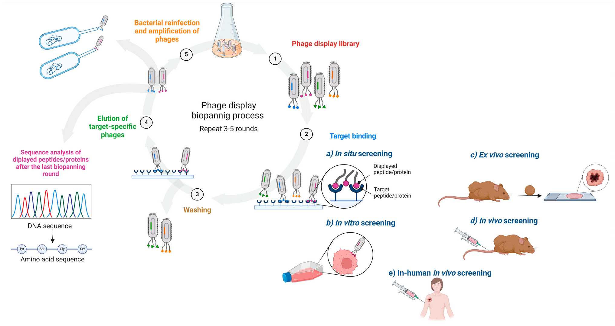 Fig. 1 Schematic of an affinity-driven process in which phage-displayed libraries are screened against a variety of targets and target-specific phage-displayed (poly)peptides (e.g., novel cancer ligands) are subsequently identified.1, 3
Rapid and Cost-Effective Methodology
Established Technique with Extensive Applications
Constraints Due to Bacterial Expression Systems
Challenges with Complex Protein Folding
Ribosome display creates a complex between mRNA, ribosome, and nascent polypeptide in vitro. Because the system is cell-free, it avoids transformation limitations and cellular toxicity.
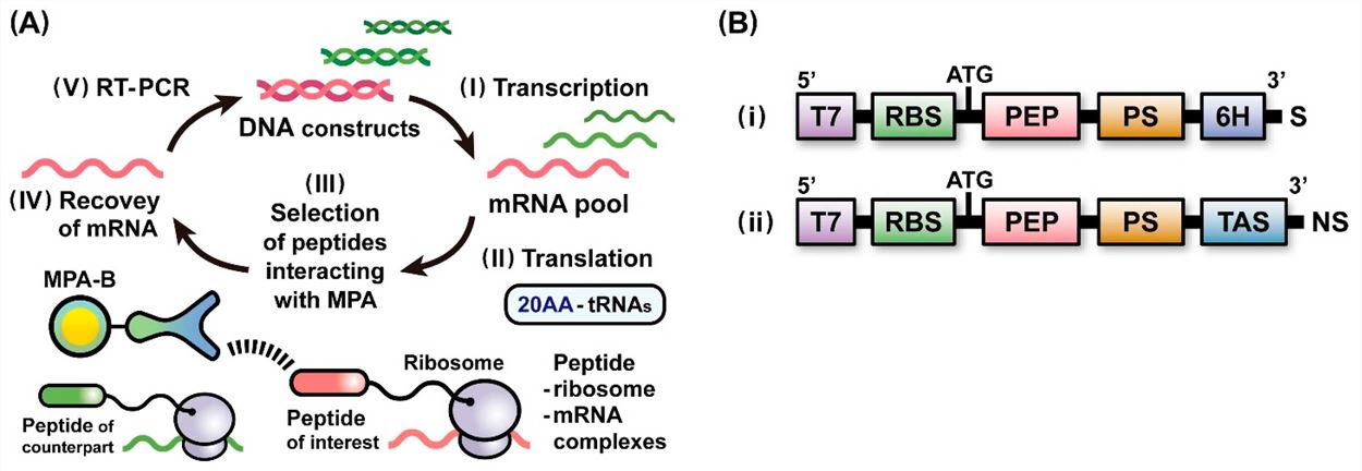 Fig. 2 Ribosome display selection for identifying the peptide of interest.2, 3
Capability to Handle Very Large Libraries
Suitable for Proteins Toxic to Cells
Stability Concerns of Complexes
Technical Challenges
Researchers choose between phage display and ribosome display technologies based on technical specifications, biological requirements, and application necessities. Molecular biologists rely on both phage display and ribosome display technologies which exhibit significant differences in their operational mechanisms and scalability as well as their suitability for various protein classes.
The primary difference between phage display and ribosome display systems centers on their capacity to store libraries.
Implication: When the highest possible library diversity is necessary during initial discovery stages or when targeting rare high-affinity variants ribosome display proves to be the perfect technique.
Implication: Ribosome display provides greater adaptability for toxic or unstable proteins which are hard to express while phage display remains beneficial when bacterial expression compatibility is adequate.
Implication: Ribosome display provides a platform to access structurally complex targets, increasing the scope of protein engineering and binder evolution projects.
Implication: Neither system is optimal for studying proteins that depend on eukaryotic PTMs. For such applications, yeast, insect, or mammalian display systems may be preferred. Nevertheless, ribosome display offers a more customizable platform for non-standard chemistry integration.
Implication: While ribosome display offers greater theoretical flexibility, phage display remains the more practical and reproducible system in many industrial workflows.
Implication: Cost-efficiency favors phage display, especially in high-throughput or early screening environments.
Table 1. Key Comparisons Between Phage Display and Ribosome Display
| Feature | Phage Display | Ribosome Display |
| Library Capacity | 10⁷–10¹⁰ | >10¹² |
| Expression System | E. coli-based | Cell-free (in vitro transcription/translation) |
| Complex Protein Compatibility | Limited | High |
| Post-Translational Modifications | Minimal support | Minimal support (modification possible) |
| Complex Stability | High (phage particles are robust) | Lower (complex is fragile) |
| Suitability for Toxic Proteins | Poor | Excellent |
| Automation/Scalability | High | Moderate |
| Cost | Low | Moderate to high |
The phage display system stands as the most reliable method in molecular display because of its straightforward application and cost-effective nature which makes it ideal for antibody research as well as peptide selection. Ribosome display supports extensive library sizes through its cell-free system flexibility which makes it ideal for directed evolution applications as well as engineering complex proteins and customized projects. Creative Biolabs technical teams understand both platforms thoroughly and help clients choose between them while optimizing or transitioning systems to meet specific research or drug development needs.
Learn more about Creative Biolabs custom phage display and ribosome display services:
All listed services and products are For Research Use Only. Do Not use in any diagnostic or therapeutic applications.








