
Creative Biolabs is offering the most comprehensive services for antibody development projects. With strict regulation and effective execution, we are dedicated to providing the most valuable solutions to complete your projects.
HighlightsCreative Biolabs is offering the most comprehensive services for antibody development projects. With strict regulation and effective execution, we are dedicated to providing the most valuable solutions to complete your projects.
HighlightsCreative Biolabs has established a broad range of platforms for developing novel antibodies or equivalents. These cutting-edge technologies enable our scientists to meet your demands from different aspects and tailor the most appropriate solution that contributes to the success of your projects.
HighlightsWith deep understanding in antibody-related realms and extensive project experience, Creative Biolabs offers a variety of references to help you learn more about our capacities and achievements, including infographic, flyer, case study, peer-reviewed publications, and all kinds of knowledge that can assist your projects. You are also welcome to contact us directly for more specific solutions.
HighlightsGet a real taste of Creative Biolabs, one of the most professional custom service providers in the world. We are committed to providing highly customized comprehensive solutions with the best quality to advance your projects.
HighlightsMolecular biology experienced a transformation with phage display technology which allows researchers to present peptides and proteins on bacteriophage surfaces. The multivalent phage display technology stands out as a powerful approach for creating high-affinity binders and refining ligand interactions among its various applications. Multivalent phage display permits multiple target molecule copies per virion which boosts avidity and selection efficiency compared to monovalent display's single-copy presentation.
Multivalent phage display involves displaying numerous copies of an external protein or peptide on a phage particle's surface. The method uses bacteriophage coat proteins including pIII, pVIII, and pIX to present exogenous molecules densely on the phage surface. The fundamental principles include:
Both multivalent and traditional monovalent phage display systems operate on the same basic principles yet they exhibit differences in binding efficiency and selection outcomes as well as practical applications. It is essential to recognize the differences between multivalent and monovalent phage display systems to select appropriate methods for antibody engineering as well as ligand screening and molecular interaction studies.
Despite its advantages, multivalent display has certain limitations that may affect experimental outcomes.
Monovalent display, in contrast to multivalent systems, presents only one or a limited number of copies of the displayed molecule per phage particle. This allows for accurate affinity ranking and precise interaction characterization.
Features of Monovalent Display:
Table. 1 Comparison with Monovalent Display Systems
| Feature | Multivalent Display | Monovalent Display |
| Binding Strength | High (due to avidity effect) | Low (single binding site) |
| Selection Efficiency | Fast (strong interactions increase retention) | Slower (single-site interactions) |
| Steric Hindrance | Moderate to high (depending on density) | Minimal |
| Conformational Fidelity | May distort some proteins | Preserves native structure |
| False Positives | More common due to non-specific interactions | Less common |
| Affinity Measurement Accuracy | Limited (multiple interactions interfere) | High (single interactions measured) |
| Best Use Case | Early-stage screening, epitope mapping, rapid selection | Affinity maturation, precise ranking, therapeutic antibodies |
Phage coat proteins provide the structural basis for multivalent display. The major and minor coat proteins facilitate the insertion of foreign peptides and proteins.
| Coat Protein | Function in Phage Display |
| pIII | Involved in phage infectivity, commonly used in antibody fragment display. |
| pVIII | Major coat protein enabling high-density display of small peptides. |
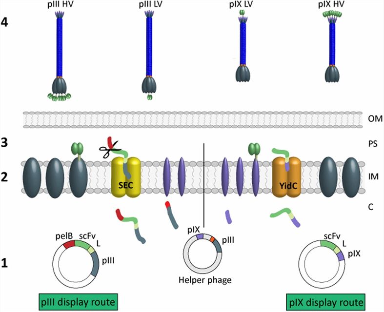 Fig. 1 Overview of pIII and pIX display routes and the four library groups.1, 3
The selection of vectors must be efficient to achieve optimal results in multivalent phage display systems. Phage display utilizes two primary vector types which include phage vectors and phagemids that possess unique characteristics. The packaging and expression of displayed proteins depend heavily on helper phages which are especially critical for phagemid-based systems.
In multivalent phage display systems phage vectors function differently from phagemids. Phage vectors allow incorporation of foreign genes into their genomes but phagemids depend on helper phages for their packaging process. The selection of these vectors determines how well display efficiency operates along with the valency and expression control capabilities.
Table 2. Comparison of Phage Vectors and Phagemids
| Feature | Phage Vector | Phagemid |
| Integration | Directly integrates foreign gene into phage genome | Requires helper phage for packaging |
| Display Valency | High (multivalent) | Low to moderate (monovalent or low multivalent) |
| Expression Control | Limited | High (inducible promoters can be used) |
| Packaging Efficiency | High | Dependent on helper phage efficiency |
| Suitability for Antibody Display | Limited | High (allows for monovalent scFv or Fab display) |
| Best Use Case | Peptide display, high-density protein display | Antibody display, controlled expression |
Helper phages provide essential packaging functions for phagemid-based display systems. They supply structural proteins required for phage particle assembly, influencing display valency and library diversity.
Table 3. Comparison of Different Helper Phages
| Helper Phage | Key Feature | Impact on Display |
| M13KO7 | Wild-type helper phage | Produces phage particles with high background wild-type coat proteins, leading to lower display efficiency |
| CM13d3 | Defective pIII gene | Selectively packages phagemid-displayed proteins, increasing display efficiency for scFv |
| Hyperphage | Lacks functional pIII | Promotes high-density multivalent display, useful for peptide library selection |
| M13cp-CT | Optimized for antibody display | Facilitates full-diversity antibody libraries, improving binding clone retrieval |
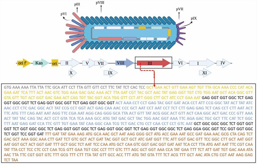 Fig. 2 Structural organization of the M13K07 phage.2, 3
Constructing a multivalent phage display library requires careful selection of vector systems, display strategies, and engineering techniques to ensure high-quality, diverse, and functionally relevant libraries. The process involves vector design, gene insertion, library amplification, and quality control. Successful library construction facilitates high-affinity ligand selection, antibody discovery, and protein-protein interaction studies.
The pIX coat protein is widely used in multivalent phage display due to its ability to tolerate foreign peptide insertions without significantly affecting phage infectivity. Designing effective pIX-based vectors requires optimization of several factors:
The construction of a high-diversity, functionally relevant phage display library requires robust cloning and selection techniques. The quality and diversity of the initial library directly influence selection success.
Ensuring library integrity and display efficiency is essential for effective biopanning and selection.
Table 4. Summary of Multivalent Phage Display Library Construction Workflow
| Step | Description | Key Considerations |
| 1. Vector Design | Engineering pIX-based vectors for multivalent display. | Optimize leader sequences, linkers, and promoters. |
| 2. DNA Library Construction | Insert diverse gene sequences into vectors. | Use error-prone PCR, DNA shuffling, or random mutagenesis. |
| 3. Transformation & Amplification | Introduce recombinant vectors into E. coli. | Maintain high-efficiency transformation for large libraries. |
| 4. Phage Rescue | Helper phage assists in packaging. | Choose helper phage to control display valency. |
| 5. Library Validation | Verify display efficiency and gene diversity. | Perform PCR, sequencing, ELISA, and Western blot. |
| 6. Biopanning & Selection | Screen library against target molecules. | Use iterative rounds for affinity maturation. |
Multivalent phage display represents a powerful and versatile technology for molecular recognition and biotechnological applications. By leveraging avid binding effects, high-throughput screening, and enhanced selection efficiency, this system has expanded the frontiers of antibody engineering, drug discovery, and targeted delivery. Creative Biolabs leads in cutting-edge multivalent phage display services. Contact us to leverage our expertise in custom antibody discovery solutions!
Learn more about Creative Biolabs phage display services:
All listed services and products are For Research Use Only. Do Not use in any diagnostic or therapeutic applications.








