iPSC-derived cardiomyocyte differentiation mimics human cardiac development to produce patient-specific cardiomyocytes, where upstream culture conditions and downstream purification processes serve as critical determinants of success. Creative Biolabs incorporates cutting-edge research insights into a GMP-compliant workflow, furnishing highly pure, fully mature cardiomyocytes through advanced RNA-switch purification and metabolic reprogramming technologies to facilitate drug discovery, disease modeling, and regenerative therapy studies.
Discover How We Can Help - Request a Consultation
iPSC-derived cardiomyocyte differentiation is a core regenerative medicine technology that guides induced pluripotent stem cells to differentiate into functional cardiomyocytes and their subtypes via in vitro induction systems mimicking embryonic cardiac development. This technology overcomes the shortcomings of primary cardiomyocytes, such as limited sources, poor in vitro proliferation, and ethical restrictions, and has broad applications in cardiovascular disease modeling, drug cardiotoxicity evaluation, and cell-based cardiac repair therapies.
The differentiation process recapitulates the in vivo embryonic cardiac development pathway, and iPSCs are induced to differentiate step by step through precise regulation of key signaling pathways and addition of cytokines/small molecules. It consists of three core stages:
| Protocol Type | Process Characteristics | Advantages | Disadvantages |
|---|---|---|---|
| Monolayer Adherent Differentiation | Induce differentiation in a 2D adherent culture system, with sequential addition of pathway regulators | Simple operation, high throughput, easy for process optimization | Low maturation degree, heterogeneous cell population, weak contractile function |
| 3D Spheroid/Organoid Differentiation | Culture iPSC-derived cardiac cells in suspension to form 3D spheroids or cardiac organoids | High cell maturity, strong functional stability, and a close to in vivo cardiac microenvironment | Complex operation, high cost, not suitable for large-scale production |
| Directed Differentiation into Cardiomyocyte Subtypes | Adjust induction factors and time windows to generate specific subtypes | Meets the needs of subtype-specific disease modeling and drug screening | Strict induction conditions, low subtype purity |
To confirm the successful generation of functional cardiomyocytes, the following validation assays are required:
The differentiation process at Creative Biolabs is a highly standardized and rigorous sequence designed for maximum yield and functional maturity.
Creative Biolabs delivers a premium, scalable, and fully customizable Cardiomyocyte Differentiation Service designed to meet the rigorous demands of global biology experts and pharmaceutical innovators. Our offering includes:
Seamless transition from laboratory-scale pilot studies to large-scale high-throughput cell production.
Bespoke optimization of culture conditions and subtype-specific patterning (atrial, ventricular, or nodal) to maximize yield for your specific application.
Utilization of high-volume industrial-grade systems ensuring consistency across batches of over 109 cells.
Integration of Quality-by-Design (QbD) and Process Analytical Techniques (PAT) to monitor cardiac maturation in real-time.
Guaranteed stability of your iPSC lines throughout the pre-cultivation and expansion phases.
Strict adherence to Hazard Analysis Critical Control Point (HACCP) approaches and GMP-certified procedures throughout the entire differentiation workflow.
High-standard quality control tools, including Patch-clamp and MEA, are used to quantify and evaluate the functional quality of every batch.
Experience the Creative Biolabs Advantage - Get a Quote Today
To assess the effect of the culture medium on the differentiation of induced pluripotent stem cells (iPSCs) into cardiomyocytes, iPSCs were pre-cultured using iMatrix511 + StemFit, LN521 + mTeSR, and LN521 + Essential 8. Cell samples were examined using techniques such as flow cytometry, qPCR, and immunofluorescence. These results indicated that distinct brown cells were observed under all culture conditions. The cardiac muscle cell marker TNNT2 mRNA was also continuously expressed in LN521 plus E8 from days 2 to 10, repeatedly inducing sustained cardiac differentiation.
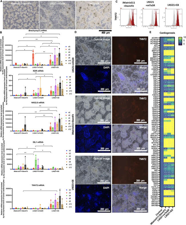 Fig.1 The influence of different component-based culture media on the potential of iPSC differentiation into cardiac cells.1
A: We go beyond simple differentiation by implementing metabolic reprogramming, switching the cells from glycolysis to fatty acid oxidation, which promotes adult-like mitochondrial bioenergetics and contractile force.
A: By using our RNA-switch technology to eliminate residual iPSCs, we consistently achieve populations that are >95% positive for cardiac markers like cTnT and α-actinin.
A: Yes, we specialize in custom differentiation. We can take your specific iPSC lines and apply our optimized protocols to generate high-quality cardiomyocytes tailored to your study.
A: Absolutely. Our xeno-free cryopreservation protocols are designed to maintain high viability. Most cultures resume spontaneous and synchronous contraction within 48 to 72 hours post-thaw.
A: While primary cells are difficult to source and have limited lifespans, our iPSC-derived cells provide a limitless, consistent, and ethically sound alternative with the added benefit of patient-specific genetic backgrounds.
Creative Biolabs offers a world-class platform for the generation, maturation, and purification of iPSC-derived cardiomyocytes. From upstream substrate optimization to advanced metabolic reprogramming and safety-critical RNA-switch purification, we provide the tools necessary to bridge the gap between benchtop discovery and clinical application.
Contact Our Team for More Information and to Discuss Your Project
Reference
For Research Use Only. Not For Clinical Use.