Human iPSC-derived neuronal differentiation transforms regenerative medicine, using pathways like Dual-SMAD/Wnt and small molecules to generate functional neurons, avoiding ethical/immunological issues as cited in recent studies. Creative Biolabs offers end-to-end, small-molecule-driven services, delivering patient-specific, fully characterized neuronal subtypes. These models enable physiologically relevant disease studies and drug screening, supported by validated products and profiles.
Discover How We Can Help - Request a Consultation
iPSC-derived neuronal cell differentiation is a core technology in regenerative medicine and neurobiology research. It reprograms somatic cells into induced pluripotent stem cells (iPSCs), then guides iPSCs to differentiate into functional neuronal cells and their subtypes through in vitro induction protocols. This technology provides a stable, patient-specific cell source for neurological disease modeling, drug screening, and potential cell replacement therapies.
The differentiation process mimics the in vivo neural development trajectory of embryonic stem cells. By precisely regulating extracellular signaling pathways (e.g., TGF-β, Wnt, Notch) and adding specific small-molecule inducers, researchers can direct iPSCs to sequentially differentiate through three key stages:
| Protocol Type | Process | Advantages | Disadvantages |
|---|---|---|---|
| Embryoid Body (EB)-Based Method | Suspend iPSCs to form 3D embryoid bodies, then culture them in neural induction medium to generate NPCs, which are then plated for terminal differentiation into neurons |
1. High differentiation efficiency 2. Simulates the in vivo embryonic development microenvironment |
1. Complex operation 2. Difficult to control the uniformity of cell differentiation |
| Monolayer Adherent Differentiation Method | Culture iPSCs as a monolayer, directly add small-molecule inhibitors (e.g., SB431542, LDN193189) to inhibit TGF-β and BMP pathways, and induce the direct differentiation of iPSCs into NPCs |
1. Simple operation 2. Good cell uniformity 3. Suitable for large-scale production |
1. Relies on high-purity iPSC monolayers 2. Strict requirements for culture conditions |
| Small-Molecule Inducer | Core Target/Mechanism | Core Function |
|---|---|---|
| SB431542 | Inhibits TGF-β/Activin/Nodal pathway | Blocks mesendodermal differentiation, promotes neuroectoderm formation |
| LDN193189 | Inhibits the BMP pathway | Synergizes with SB431542 to improve induction efficiency |
| RA (Retinoic Acid) | Binds to retinoic acid receptor (RAR) | Regulates axial differentiation, determines neuronal spatial fate |
| SAG | Activates the Shh pathway | Regulates ventral neuronal fate, specifies subtypes |
| BDNF | Binds to the TrkB receptor | Promotes neuronal survival, maturation, and synaptogenesis |
To initiate a project, clients typically provide Starting Materials such as:
Experience the Creative Biolabs Advantage - Get a Quote Today
At Creative Biolabs, we recognize that every neurological research project possesses unique requirements. We provide highly flexible and customized Functional Neuronal Cell Differentiation Service tailored to your specific experimental goals:
Tailored differentiation protocols for a wide array of subtypes, including dopaminergic, motor, sensory, and cortical neurons, specifically designed to meet your project's phenotypic requirements.
Utilization of advanced, chemically defined small-molecule cocktails to ensure rapid, stable, and highly reproducible neurogenesis.
Capability to handle projects ranging from laboratory-scale pilot studies to large-scale industrial batches for high-throughput screening (HTS).
Integration of Multi-Electrode Array (MEA) and calcium imaging to guarantee that delivered neurons exhibit mature electrophysiological profiles.
Development and maintenance of patient-derived iPSC lines with confirmed genetic backgrounds (e.g., specific mutations for PD, AD, or ALS).
To track the development and maturation of human cortical neurons in vitro, we utilized a previously established iPSC line. Via a dual inhibitor approach, the iPSCs were differentiated into forebrain neurons, astrocytes, and neuronal precursor cells (NPCs). The S11 NPCs were then induced to differentiate into human neurons in a neuronal differentiation medium supplemented with BDNF (10 ng/mL), GDNF (10 ng/mL), and dibutyryl cyclic adenosine monophosphate (1 mM), followed by in vitro culture for 10 weeks. Neuron-specific markers βIII-tubulin and MAP2 were detectable as early as the first week of culture.
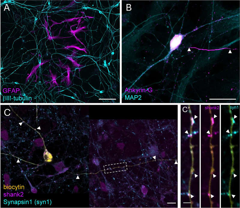 Fig.1 The differentiated h-iPSC-N cultures expressed neuronal and glial cell markers in the first week after maturation.1
A: We utilize a multifactorial validation approach. Beyond simple marker expression (ICC), we employ a Multi-Electrode Array (MEA) to monitor spontaneous firing and network synchronization. This confirms that the neurons are electrophysiologically active and capable of synaptic communication.
A: Yes. We specialize in custom differentiation. If you provide the specific iPSC line or require us to use CRISPR/Cas9 to introduce a mutation, we can tailor the patterning and maturation protocol to suit the expected phenotypic expression of that specific rare disease.
A: Small molecules are more cost-effective, offer better penetration into 3D structures like organoids, and provide more stable, rapid, and reversible biological effects. This leads to higher reproducibility compared to protein-based induction.
A: Yes, we have extensive experience integrating our differentiated neurons into 3D organoids and microfluidic devices to mimic the complex architecture of the human blood-brain barrier and neural circuits.
A: We implement a rigorous "Negative Selection" protocol, removing any cells expressing pluripotency markers like SSEA-4. This is followed by in vivo teratoma formation assays for any batches intended for clinical safety studies.
Creative Biolabs offers a robust and scalable Functional Neuronal Cell Differentiation Service designed to meet the rigorous demands of modern drug discovery and regenerative medicine. From providing high-purity specialized neuronal subtypes to delivering functional validation data, we are committed to accelerating your path to the clinic.
Contact Our Team for More Information and to Discuss Your Project
Reference
For Research Use Only. Not For Clinical Use.