Musculoskeletal Cell Differentiation enables the generation of high-purity bone tissue via TGF-β pathway modulation and molecular brake inhibition, offering superior scalability for bone disease modeling and regenerative therapeutics research. Creative Biolabs delivers functionally validated osteoblasts and osteocytes with high transcriptomic fidelity via advanced reprogramming and 3D platforms, streamlining orthopedic drug discovery and tissue engineering projects.
Discover How We Can Help - Request a Consultation
iPSC-derived musculoskeletal cell differentiation refers to the in vitro directional induction of induced pluripotent stem cells into functional osteoblasts, osteocytes, and other bone lineage cells by simulating the process of embryonic osteogenesis. This technology circumvents the limitations of traditional bone tissue engineering seed cells (such as limited sources, immune rejection, and ethical disputes) and has broad application prospects in bone defect repair, osteoporosis disease modeling, and drug screening.
The process recapitulates the two pathways of embryonic osteogenesis: intramembranous ossification and endochondral ossification, and is generally divided into three core stages:
| Protocol Type | Operation Characteristics | Advantages | Disadvantages |
|---|---|---|---|
| 2D Adherent Induction | Induce differentiation on tissue culture plates with osteogenic medium | Simple operation, low cost, easy to observe cell morphology changes | Low degree of cell mineralization, poor simulation of the in vitro bone microenvironment |
| 3D Scaffold Culture | Seed iPSC-derived MSCs onto biomaterial scaffolds (e.g., hydroxyapatite, collagen scaffolds) for osteogenic induction | Good cell-matrix interaction, high mineralization efficiency, closer to in vivo bone tissue structure | Complex scaffold preparation, high cost |
| Co-culture System | Co-culture iPSC-derived cells with endothelial cells or chondrocytes | Promotes vascularization and osteogenic differentiation synergy, improving the survival rate of engineered bone in vivo | Complex system design, difficult to control the proportion of co-cultured cells |
Our standardized yet flexible workflow ensures that every project meets the rigorous demands of biopharmaceutical research.
At Creative Biolabs, we go beyond simple cell delivery. We provide a comprehensive, industrial-grade solution for Musculoskeletal Cell Differentiation Service tailored to the precise needs of biology experts and pharmaceutical innovators.
From laboratory-scale pilot studies to large-scale industrial cell production.
Advanced codon and signaling optimization to maximize the expression of osteogenic markers in selected iPSC lines.
High-capacity 3D bioreactor systems and specialized incubation environments to ensure bulk-order consistency.
Rigorous assessment of genomic stability in cell banks and throughout long-term osteogenic maturation phases.
Implementation of Quality-by-Design (QbD) and Process Analytical Techniques (PAT) for every batch.
We follow strict Hazard Analysis Critical Control Point (HACCP) approaches and Good Manufacturing Practice (GMP) principles to ensure aseptic and high-quality production.
High-standard quality control tools, including scRNA-seq, to quantify and evaluate the biological quality and lineage purity of the products.
Experience the Creative Biolabs Advantage - Get a Quote Today
To induce the differentiation of iPSCs into osteoblasts, researchers conducted osteogenic induction of hiPSCs on Matrigel-coated culture dishes within type I collagen gels. Vertical sections harvested at sequential time points during induction revealed a gradual increase in cell density inside the gels. Horizontal sections obtained on day 14 showed that the gel surface was covered by a sheet of cuboidal cells, while cells embedded in the gel exhibited a dendritic morphology and formed connections with adjacent cells. Immunostaining of vertical sections confirmed positive expression of DMP1 in intra-gel cells, whereas immunostaining of horizontal sections demonstrated a reticular distribution of human type I collagen (COL I) beneath the cuboidal cell layer.
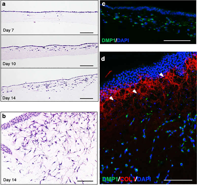 Fig.1 Induction of osteoblastic and osteocytic cells from hiPSCs (414C2) on type I collagen gel.1
A: We utilize standardized, xeno-free differentiation protocols that have been validated across dozens of healthy and disease-specific lines. Before starting, we conduct a pluripotency screen to ensure your specific line is "differentiation-ready," mitigating the risk of lineage bias.
A: Yes, our iPSC-derived osteogenic progenitors are specifically optimized for integration with various bio-inks. Their high proliferative capacity makes them ideal for the high cell densities required for successful 3D bioprinting of functional bone scaffolds.
A: Our 2D service provides rapid, high-purity osteoblasts for initial screening. Our 3D service uses Type I collagen gels to promote gel invasion and terminal maturation into osteocytes, which is essential if your research focuses on bone mechanotransduction or terminal homeostasis.
A: Yes. Given the paracrine nature of bone repair, we offer specialized characterization of Extracellular Vesicles (EVs) produced during the differentiation process, including NTA size analysis and miRNA cargo profiling.
A: Unlike MSCs, which suffer from donor-to-donor variability and "replicative senescence" (loss of potency over time), our iPSC-derived cells provide an infinite, standardized supply with higher genetic consistency, allowing for more reproducible long-term studies.
Creative Biolabs offers a comprehensive suite of iPSC differentiation services, characterization assays, and secretome engineering solutions to support your most ambitious orthopedic projects. Our team of experts is ready to discuss your specific project needs and provide a tailored solution that accelerates your path to clinical discovery.
Contact Our Team for More Information and to Discuss Your Project
Reference
For Research Use Only. Not For Clinical Use.