Embryoid Body Formation & Characterization Service
IntroductionEmbryoid Body Formation & CharacterizationWhat We Can OfferCustomer ReviewsFAQsRelated Sections
Introduction
Creative Biolabs' Embryoid Body Formation & Characterization Service delivers high-quality 3D EBs that recapitulate early embryogenesis and enable robust tri-germ layer differentiation. These validated EBs serve as reliable building blocks for drug discovery, disease modeling, and pluripotency assessment. Our automated systems and molecular fingerprinting ensure standardization and reproducibility, streamlining your biopharmaceutical pipeline development.
Discover How We Can Help - Request a Consultation
Embryoid Body Formation & Characterization
Embryoid body (EB) formation is a classic in vitro assay for evaluating iPSC pluripotency. When undifferentiated iPSCs are cultured in suspension without matrix adherence and pluripotency-maintaining factors, they aggregate into spherical EBs that spontaneously differentiate into cells of all three germ layers, mimicking early embryonic development.
Key Experimental Procedures
Cell Preparation
Harvest undifferentiated iPSC colonies in a single-cell or small-cluster suspension using enzymatic or mechanical dissociation methods. Ensure cell viability to support efficient EB formation.
Suspension Culture for EB Formation
Seed the iPSC suspension into ultra-low attachment culture plates to prevent cell adhesion. Culture in EB induction medium (usually serum-containing or defined medium without leukemia inhibitory factor, LIF). Incubate for 3-7 days, with medium changes every 1-2 days, to allow the formation of compact, spherical EBs.
EB Maturation (Optional)
For further differentiation, transfer the formed EBs to adherent culture plates. The cells will migrate out of the EBs and adhere to the substrate, continuing to differentiate into specialized cell types over 2-4 weeks.
EB Characterization
Morphological observation: Monitor EB size, shape, and integrity under a light microscope during culture.
Germ layer marker detection: Verify the presence of three germ layer derivatives using immunofluorescence staining, RT-qPCR, or flow cytometry. Typical markers include PAX6 (ectoderm), Brachyury (mesoderm), and SOX17 (endoderm).
Core Detection Methods for EBs
EB characterization focuses on morphological features and tri-germ layer differentiation potential, with specific methods as follows:
Monitor the EB formation process and morphological traits dynamically using an inverted microscope. Record parameters including size, sphericity, edge regularity, and internal structural uniformity, while checking for adhesion, fragmentation, or core necrosis.
This method enables rapid preliminary evaluation of EB formation quality with simple operation and no need for special reagents.
Molecular-Level Detection (Verification of Germ Layer Markers)
RT-qPCR/Quantitative Real-Time PCR: Quantify transcriptional levels of ectoderm, mesoderm, and endoderm marker genes in EBs to assess tri-germ layer differentiation balance.
Immunofluorescence Staining/Immunocytochemistry: Localize and visualize tri-germ layer marker proteins; observe positive cell ratio and distribution to confirm multi-lineage differentiation capacity.
Flow Cytometry: Stain dissociated EB single-cell suspensions with specific markers for high-throughput quantitative analysis of positive cell proportions across three germ layers.
Functional-Level Detection (Evaluation of Directed Differentiation Potential)
Seed EBs onto adherent culture dishes and culture them under conditions without exogenous inducers or with specific induction protocols. Observe whether migrated cells can differentiate into specialized functional cell types, such as neurons (ectoderm), cardiomyocytes (mesoderm), and hepatocyte-like cells (endoderm). Verify the differentiation potential of EBs through functional validation.
Criteria for High-Quality EBs
High-quality EBs must meet two core requirements: morphological integrity and balanced differentiation potential.
Morphological Criteria
Uniform size: The diameter is typically within the range of 100-300 μm, with minimal variation among individual EBs in the same batch and no abnormally large or small ones.
Regular morphology: Present as compact spherical structures with smooth, rounded edges, without obvious indentations, fragmentation, or cell clumping.
Healthy internal structure: No significant core necrosis is observed under microscopy, with uniform cell distribution and consistent light transmittance.
Stable formation efficiency: After 3-7 days of suspension culture, the EB formation rate is ≥ 80%, with stable morphology and no tendency to disintegrate easily.
Differentiation Potential Criteria
Balanced tri-germ layer differentiation capacity: Molecular assays confirm robust expression of markers across all three germ layers, with no biased differentiation toward any single lineage.
Normal function of differentiated cells: Adherent culture yields cells with typical morphology and function, including cardiomyocyte spontaneous beating and neuronal synapse-like structure formation.
What We Can Offer
At Creative Biolabs, we provide a robust and highly adaptable platform for Embryoid Body Formation & Characterization Service, tailored to the exacting standards of biology experts. Our capabilities include:
One-stop 3D culture service
from laboratory-scale pilot studies to large-scale industrial differentiation.
Efficient upstream and downstream process development
ensuring seamless transition from EB formation to specialized organoid maturation.
Scalable high-throughput aggregation
using robotic systems and multi-well plate platforms for thousands of uniform aggregates.
Well-established quality systems
incorporating Quality-by-Design (QbD) principles and process analytical techniques (PAT) for every batch.
Customized differentiation protocols
where culture conditions, media additives, and aggregation modes (batch or continuous) are optimized to maximize your specific lineage yield.
Strict aseptic verification
and GMP-compliant procedures throughout the differentiation and characterization process.
High-standard quality control tools
including scRNA-seq, qPCR, and advanced histology to quantify and evaluate germ-layer commitment.
Documentation and traceability
where the strain origin and iPSC reprogramming history are assessed and approved by our qualified quality assurance team.
Experience the Creative Biolabs Advantage - Get a Quote Today
Case Study
A production platform based on air microfluidics (IAMF) enables ultra-high-throughput preparation of homogeneous and functional pluripotent stem cell-derived embryoid bodies (EBs) and spheres. Researchers loaded approximately 20 human pluripotent stem cells (hPSCs) into microcapsules and conducted suspension culture in Essential 8 (E8) stem cell medium. The microencapsulated hPSCs aggregated to form dense 3D hPSC spheres within two days. The pluripotency of these spheres was confirmed via the expression of pluripotency markers Sox2 and Oct3/4, with levels comparable to those observed in traditional monolayer cultures.
Fig.1 Embryonic bodies were constructed using microcapsules with controllable size.1
Customer Reviews
FAQs
Q: What is the primary advantage of EB formation over 2D differentiation?
A: EBs provide a 3D microenvironment that allows for natural cell-to-cell signaling and spatial organization, which is essential for developing complex tissues like the neural retina.
Q: Do you offer custom media formulations during EB formation?
A: Yes, we provide flexible, custom induction protocols to meet the specific requirements of your target lineage.
Q: How do you ensure the EBs are free from genomic abnormalities?
A: We perform routine karyotyping and G-banding, alongside qPCR to monitor for common reprogramming-induced mutations.
Q: Can your EBs be used for drug toxicity screening?
A: Absolutely. Our standardized EBs are specifically designed for HTS applications, providing reproducible data for toxicology and efficacy studies.
Q: What is the difference between your 'forced' and 'non-forced' aggregation?
A: Forced aggregation (SCP) uses centrifugation or V-bottom plates for high uniformity, while non-forced (CP) relies on natural sedimentation, which some researchers prefer for maintaining delicate cell-surface markers.
Creative Biolabs offers an end-to-end solution for your 3D culture needs, from initial iPSC reprogramming to advanced functional validation. Our platform ensures that your research is supported by the highest standards of biological accuracy and industrial scalability.
Contact Our Team for More Information and to Discuss Your Project
Related Sections
Reference
van Loo, Bas, et al. "Mass production of lumenogenic human embryoid bodies and functional cardiospheres using in-air-generated microcapsules." Nature communications 14.1 (2023): 6685. https://doi.org/10.1038/s41467-023-42297-0. Distributed under Open Access license CC BY 4.0, without modification.
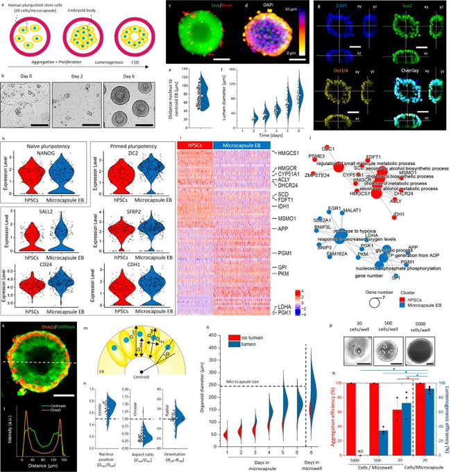 Fig.1 Embryonic bodies were constructed using microcapsules with controllable size.1