Creative Biolabs' Teratoma Formation Assay Service is the definitive functional test for iPSC pluripotency, validating multi-lineage differentiation that in vitro assays cannot replicate by proving response to systemic developmental signals and epigenetic remodeling. Leveraging advanced xenograft platforms, standardized histological analysis, and immunodeficient mouse models, we deliver quantitative data, expert pathological review, and gold-standard evidence for regulatory filings and high-impact publications.
Discover How We Can Help - Request a Consultation
The teratoma formation assay, the gold standard functional test for iPSC pluripotency, is essential for iPSC research and application for three core reasons. It verifies functional pluripotency by confirming iPSCs' ability to differentiate into all three germ layers in vivo, a criterion that in vitro assays cannot fully reflect. It ensures safety by eliminating risky cell lines and provides a quality control basis for iPSC culture and modification to maintain cell quality stability.
When undifferentiated iPSCs are transplanted into immunodeficient mice (e.g., NOD/SCID mice), they form teratomas, benign tumors containing heterogeneous tissues derived from the three germ layers. The presence of these diverse tissues directly verifies the pluripotent potential of iPSCs.
To ensure the most accurate assessment of pluripotency, Creative Biolabs utilizes a rigorous, multi-step workflow optimized for reproducibility and sensitivity.
As an industry leader in regenerative medicine services, Creative Biolabs provides a comprehensive and fully customizable platform for Teratoma Formation Assay Service. We understand that every iPSC project has unique requirements, and we offer bespoke solutions that bridge the gap between bench-side research and clinical application.
tailored to specific cell line growth characteristics and client-defined endpoints;
(NSG, Nude, SCID) to maximize engraftment success;
across various anatomical sites, including subcutaneous, intramuscular, and subcapsular injections;
and proprietary scaffold optimizations to enhance sensitivity for challenging cell lines;
utilizing both standard H&E and specialized IHC markers for germ-layer identification;
throughout the longitudinal in vivo study;
compliant with high-standard regulatory and peer-reviewed publication requirements.
Experience the Creative Biolabs Advantage - Get a Quote Today
Teratoma formed by human embryonic stem cell lines (such as the H9 lineage) implanted in immunodeficient mice can be stained with hematoxylin-eosin, Wechsler staining, alizarin red, Masson tricolor staining, and Wegherter's staining, which can be used to evaluate cell pluripotency.
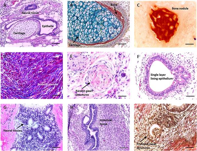 Fig.1 Morphological detection of teratoma derived from human pluripotent stem cells.1
A: While in vitro tests are faster, they only measure mRNA or surface markers. The teratoma assay is the only test that proves your cells can physically organize into complex tissues, which is a key requirement for therapeutic safety and efficacy.
A: The intramuscular or subcutaneous sites are most common for ease of monitoring. However, the kidney capsule is often used for higher sensitivity if the cell line shows poor growth characteristics in other sites.
A: We exclusively use highly immunodeficient strains like NSG or Nude mice, which lack T, B, and sometimes NK cell function, ensuring high engraftment rates for human iPSCs.
A: Yes. Beyond pluripotency, our pathologists specifically look for signs of teratocarcinoma or undifferentiated clusters, which can indicate genomic instability or incomplete differentiation.
A: Standard protocols usually involve 3-5 mice per cell line to ensure statistical significance and to account for any biological variability in tumor growth.

Creative Biolabs offers a robust, end-to-end solution for Teratoma Formation Assay Service, providing the critical validation needed for both basic research and clinical translation. Our combination of surgical precision, high-grade animal models, and expert pathology ensures that your pluripotency data is beyond reproach.
Contact Our Team for More Information and to Discuss Your Project
Reference
For Research Use Only. Not For Clinical Use.