Identifying AHCTF1 as a prognostic biomarker for aggressive tumor phenotypes. Our analysis helps evaluate its role in aberrant nuclear transport in cancer cells, where the disruption of nucleocytoplasmic trafficking can lead to the sequestration of tumor suppressors or the over-activation of oncogenic transcription factors.
Are you currently facing challenges in identifying reliable biomarkers for cell cycle progression or struggling with the detection of complex nucleoporin proteins in oncological research? Our AHCTF1 Analysis Services help you unlock the full potential of your drug discovery and diagnostic projects through Creative Biolabs' innovative high-affinity antibody development and ultrasensitive singleplex biomarker detection platforms.
Contact our team to get an inquiry now!
Introduction of AHCTF1 Analysis
AHCTF1 (AT-hook containing transcription factor 1) is a critical multifunctional protein primarily localized at the nuclear envelope. It plays a pivotal role as a scaffolding element for the Nup107–160 complex, which is essential for the assembly of the NPC during both post-mitotic nuclear envelope reformation and interphase. Structurally, AHCTF1 contains a signature AT-hook domain that facilitates its high-affinity binding to AT-rich DNA sequences on chromatin. This interaction is not merely structural; it acts as a critical "seeding point" for nuclear pore construction, effectively anchoring the pore assembly machinery to the genome at the end of mitosis to ensure the proper compartmentalization of the daughter nuclei.
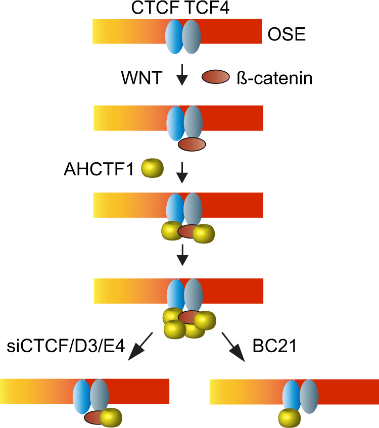 Fig.1 AHCTF1 involved in WNT/ß-catenin-AHCTF1-CTCF-eRNA signaling pathways.1
Recent literature highlights the profound significance of AHCTF1 in maintaining genomic integrity across multiple cell generations. Conclusion of contemporary studies indicates that AHCTF1 depletion or dysfunction leads to severe mitotic defects, including chronic chromosome missegregation and impaired nuclear envelope integrity, which can trigger micronuclei formation and subsequent DNA damage. Furthermore, AHCTF1 has been implicated in various signaling pathways governing cell proliferation and is frequently found significantly overexpressed in several malignancies, such as triple-negative breast cancer and certain leukemias. This overexpression often correlates with increased metabolic demand and rapid cell division, positioning AHCTF1 as a promising candidate for both diagnostic profiling and targeted therapeutic intervention.
Application of AHCTF1 Analysis
AHCTF1 analysis is instrumental across several high-impact research areas, providing deep insights into cellular architecture and pathology:
Oncology Research
Cell Cycle Studies
Investigating the intricate kinetics of NPC assembly and the precise recruitment of the Nup107–160 complex during the transition from telophase to G1. This includes analyzing how AHCTF1 coordinates with other nucleoporins to ensure the timely reformation of a functional nuclear barrier.
Drug Screening
Utilizing AHCTF1 expression levels as a high-fidelity readout for testing the efficacy of novel cell cycle inhibitors or nuclear transport modifiers. Researchers can monitor how lead compounds impact AHCTF1 localization as a proxy for their ability to disrupt mitotic progression in hyper-proliferative cells.
Developmental Biology
Mapping the spatiotemporal expression patterns of AHCTF1 during embryonic development to understand its role in tissue morphogenesis. Given its role in nuclear organization, AHCTF1 analysis can reveal how specialized cells manage nuclear envelope demands during rapid differentiation phases.
Genomic Stability Assays
Assessing how AHCTF1-chromatin interactions prevent catastrophic DNA damage and maintain the complex 3D architecture of the chromosome. This involves studying the consequences of disrupted chromatin anchoring on global gene expression and long-term genomic stability.
Service Highlights
Creative Biolabs offers a best-in-class AHCTF1 analysis platform characterized by technical excellence and reliability.
- One-Stop Service: We provide an integrated pipeline ranging from antigen design and gene synthesis to the final production of high-affinity antibodies and analytical validation.
- Diverse Antibody Formats: Our platform supports the production of full-length IgGs, scFv, Fab, and bispecific formats to suit specific assay requirements.
- Guaranteed High Purity: Utilizing advanced chromatography, we ensure purity up to 99% with ultra-low endotoxin levels, critical for sensitive diagnostic assays.
- Comprehensive QC Analysis: Every project undergoes Published Data validation, including SDS-PAGE, HPLC, and ELISA, ensuring batch-to-batch stability and specificity.
- First-in-Class Technology: Powered by our proprietary phage display and hybridoma technologies, we deliver antibodies that outperform standard commercially available reagents.
- Dedicated After-Sale Support: Our PhD-level scientists provide guidance on assay optimization to ensure optimal performance in your laboratory environment.
Service Workflow
01Initial Consultation and Project Design
Our team reviews your specific AHCTF1 research goals to customize the antibody valency and assay format (e.g., IHC, WB, or Sandwich ELISA).
02Antibody Development and Validation
Leveraging our high-throughput platform, we generate lead antibody candidates and validate them against AHCTF1 targets using Published Data to confirm affinity (KD) and specificity.
03Sample Processing and Quality Control
Upon receipt, samples undergo stringent QC to verify integrity, ensuring that the AHCTF1 protein within the matrix is not degraded.
04Data Acquisition and Analysis
Using ultrasensitive detection systems, we quantify AHCTF1 levels. Data is interpreted using advanced statistical models to provide meaningful biological context.
05Final Reporting
A detailed report containing raw data, methodology, and expert interpretation is provided to facilitate your downstream applications.
FAQs
-
How does the AT-hook domain of AHCTF1 influence its recruitment to the chromatin surface?
The AT-hook domain is a specialized DNA-binding motif that recognizes the minor groove of AT-rich sequences. In the context of mitosis, this domain allows AHCTF1 to act as a trans-acting factor that anchors the Nup107-160 complex directly to the chromosome surface. This interaction is the biochemical prerequisite for post-mitotic nuclear pore complex (NPC) assembly and ensures that nuclear envelope reformation occurs around the condensed genome.
-
What biological implications arise from the differential distribution of AHCTF1 between the nuclear envelope and the nucleoplasm?
AHCTF1 primarily functions as a transmembrane-linked scaffold at the nuclear pore, but a soluble nucleoplasmic pool also exists. Shifting ratios between these fractions can indicate changes in nuclear transport capacity or cellular stress. In diseased states, such as malignant transformation, an increase in nucleoplasmic AHCTF1 may correlate with aberrant signaling or hyper-active NPC biogenesis required for rapid cell proliferation.
-
Can current analytical techniques detect AHCTF1 at the picogram level in early-stage disease models?
Yes. Advances in ultrasensitive immunoassay technology and signal amplification allow for the detection of AHCTF1 at picogram-per-milliliter concentrations. This level of sensitivity is essential for identifying early molecular markers of genomic instability or nuclear envelope defects before macroscopic cellular changes become evident in oncological research.
-
Which experimental precautions are critical to prevent the degradation of the AHCTF1 protein during nuclear fractionation?
AHCTF1 is sensitive to proteolysis and mechanical shearing due to its large size and association with the chromatin-nuclear envelope interface. Maintaining structural integrity requires the use of isotonic buffers, a comprehensive cocktail of protease inhibitors, and strictly controlled temperatures (4℃). Furthermore, the choice of detergent is critical; non-ionic detergents are typically preferred to preserve the protein-chromatin complexes during initial lysis.
-
Why is epitope specificity more critical for AHCTF1 than for smaller, cytosolic proteins in quantitative assays?
Due to its complex multi-domain structure and extensive involvement in protein-protein interactions (such as with the Nup107-160 complex), certain epitopes of AHCTF1 may be sterically hindered or masked in its native state. High-specificity detection reagents must be validated to recognize epitopes that remain accessible under various physiological conditions, ensuring that quantitative data reflects the true protein concentration rather than fluctuations in epitope availability.
Creative Biolabs is committed to advancing the field of nuclear biology and oncology through our specialized AHCTF1 Analysis Services. By combining state-of-the-art antibody engineering with rigorous analytical validation, we empower our clients to achieve breakthroughs in biomarker discovery and drug development.
Reference
- Chachoua, Ilyas et al. "Canonical WNT signaling-dependent gating of MYC requires a noncanonical CTCF function at a distal binding site." Nature communications vol. 13,1 204. 11 Jan. 2022, Distributed under Open Access license CC BY 4.0, without modification. https://doi.org/10.1038/s41467-021-27868-3
For Research Use Only.