Comprehensive quantitative and qualitative screening for autoantibodies in serum or CSF using IHC and confirmatory immunoblotting for precise clinical diagnostics.
Creative Biolabs guarantees the highest discovery power for maximum outcomes through our premier singleplex biomarker analysis platform. We provide comprehensive amphiphysin (AMPH) analysis services, offering a variety of strategic options to help customers determine precise biomarker expression levels, mitigate developmental risks, and evaluate drug efficacy and safety with expert-led validation and sample analysis.
Introduction of AMPH
AMPH is a 125 kDa synaptic vesicle-associated protein that regulates clathrin-mediated endocytosis and vesicle release in excitable cells. Expressed primarily in neurons, neuroendocrine cells, and spermatocytes, it serves as a critical scaffold. Its modular architecture, featuring N-BAR and SH3 domains, allows multi-site phosphorylation to facilitate actin polymerization during phagocytosis, neurite outgrowth, and spermatid release from sertoli cells.
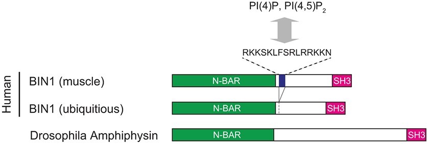 Fig.1 Human and Drosophila amphiphysin.1
Clinical research identifies AMPH antibodies as biomarkers for breast cancer, small-cell lung cancers (SCLC), and neurological disorders. Specifically, anti-AMPH autoantibodies (AMPH-IgG) are linked to paraneoplastic syndromes like Limbic Encephalitis (LE) and Stiff-Person Syndrome (SPS). In these cases, AMPH acts as an "onconeural" antigen, making its analysis vital for early cancer detection and therapeutic monitoring.
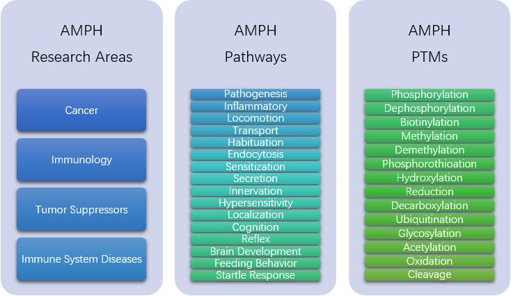
AMPH Analysis Services at Creative Biolabs
All AMPH reagents utilized in our laboratory are produced in-house under rigorous quality control to ensure consistency. With deep expertise in AMPH-IgG and associated pathologies, we provide reliable, multi-dimensional biomarker analysis. Utilizing native samples or recombinant proteins, we characterize AMPH-IgG via immunohistochemistry and confirm specificities through immunoblotting, the gold standard for identifying intracellular antibodies, ensuring absolute research accuracy.
Our featured AMPH services include, but are not limited to:
AMPH-IgG Detection
AMPH Interaction Analysis
Utilizing SPR or ITC to map protein-protein interactions (PPI) between the AMPH SH3 domain and partners like Dynamin or Synaptojanin, elucidating complex signaling.
Knockdown of AMPH
Targeted target validation using RNAi or other gene editing systems to study the functional impact of AMPH deficiency in diverse disease-relevant cell models.
AMPH PTMs Detection
Specialized mass spectrometry workflows to identify post-translational modifications (PTMs), such as phosphorylation or ubiquitination, which regulate the protein's activity and localization.
Anti-AMPH Antibody Development
Custom generation of high-affinity monoclonal or polyclonal antibodies specifically tailored for western blotting, ELISA, and advanced immunohistochemical research applications.
Service Workflow
Our AMPH Analysis follows a systematic, expert-led workflow designed for maximum precision:
01Initial Consultation
We conduct a technical review to define specific diagnostic or research goals and establish the appropriate AMPH analysis parameters.
02Sample Submission
Clients provide starting materials, such as serum, cerebrospinal fluid (CSF), tissue biopsies, or cell lysates, following our stabilization guidelines.
03Platform Execution
Our experts perform the requested services, ranging from IHC autoantibody screening on rat brain sections to high-sensitivity mass spectrometry for PTM detection.
04Verification & Quality Control
Every step is cross-validated against our in-house recombinant AMPH standards to ensure data integrity and specificity.
05Data Integration
We compile high-resolution raw data and interpreted results into a cohesive analytical framework.
06Final Delivery
A comprehensive report detailing the AMPH biomarker profile is delivered to support critical research decisions and program milestones.
Applications
Paraneoplastic Syndrome Diagnosis
Detection of anti-AMPH autoantibodies facilitates the early diagnosis of paraneoplastic neurological syndromes. This analysis acts as a critical indicator for underlying occult malignancies, particularly breast adenocarcinoma and small-cell lung cancer, often preceding radiographic tumor detection.
Synaptic Vesicle Research
Analysis of AMPH-mediated endocytosis provides insights into synaptic vesicle recycling and neurotransmitter release mechanisms. This is essential for researchers studying synaptic plasticity, neurotransmission efficiency, and the molecular basis of neurodegenerative diseases.
Target Validation for Oncology
Evaluating AMPH expression levels and its role in actin polymerization supports the validation of AMPH as a potential target in cancer research. Understanding its involvement in neurite-like outgrowth and cell migration is pivotal for developing anti-metastatic therapeutics.
Drug Mechanism of Action (MoA) Studies
AMPH Analysis helps characterize how candidate drugs influence membrane remodeling and intracellular trafficking. By monitoring AMPH phosphorylation or its interaction with Dynamin, researchers can quantify drug-induced changes in cellular endocytic flux.
Spermatogenesis and Fertility Research
Studies focusing on spermatid release and Sertoli cell function utilize AMPH analysis to investigate the cytoskeletal rearrangements required for male fertility. This application supports the development of reproductive health diagnostics and treatments.
Biomarker Discovery in Neuroendocrinology
Quantifying AMPH levels in neuroendocrine tissues serves as a specialized tool for identifying markers of cellular stress or secretion disorders. This assists in characterizing the secretory pathways of hormone-producing cells under various pathological conditions.
Service Highlights
- Gold-Standard Confirmation: We employ a dual-platform approach using Immunohistochemistry (IHC) followed by Immunoblotting to eliminate false positives in AMPH-IgG detection.
- In-house Reagent Control: Total control over the production of recombinant AMPH proteins and antibodies ensures high sensitivity and superior reagent purity.
- Isoform-Specific Pipelines: Specialized protocols are available to distinguish between neuronal AMPH1 and the muscle/brain-specific AMPH2 (BIN1) isoforms.
- High-Throughput Capability: Our platform supports large-scale screening of chemical libraries for compounds that modulate AMPH-mediated protein interactions.
- Universal Reporting: Reports integrate biochemical, biophysical, and phenotypic data, providing a holistic view of the AMPH biomarker's status.
FAQs
-
Q: What is the primary advantage of using immunoblotting over ELISA for AMPH-IgG detection?
A: Immunoblotting is considered the gold standard for intracellular antibodies like AMPH because it allows for the definitive identification of the 128 kDa protein band. Unlike ELISA, which may suffer from non-specific binding, immunoblotting ensures the antibody is reacting specifically with the target protein structure, significantly reducing the risk of false-positive results.
-
Q: Can your AMPH Analysis service distinguish between AMPH1 and BIN1 (AMPH2)?
A: Yes, our platform utilizes specific recombinant proteins and isoform-selective antibodies to differentiate between the 128 kDa AMPH1 and the various isoforms of BIN1. This distinction is critical as AMPH1 is primarily associated with paraneoplastic syndromes, while BIN1 has specific roles in muscle T-tubule formation and Alzheimer's disease progression.
-
Q: Which biological samples are most suitable for detecting anti-AMPH autoantibodies?
A: We recommend providing paired serum and cerebrospinal fluid (CSF) samples for the most comprehensive analysis. While serum is often sufficient for initial screening, CSF testing provides higher specificity for central nervous system involvement and is mandatory for confirming paraneoplastic neurological syndromes.
-
Q: How do you ensure the stability of AMPH PTMs during the analysis process?
A: We utilize specialized lysis buffers enriched with phosphatase and protease inhibitors to "freeze" the modification state of the AMPH protein. Our sample preparation protocols are optimized to prevent the degradation of labile modifications like phosphorylation, ensuring the data reflects the true physiological state.
-
Q: Is it possible to perform AMPH interaction analysis with small molecule libraries?
A: Our platform is fully equipped for high-throughput screening using Surface Plasmon Resonance (SPR). We can immobilize the AMPH SH3 domain or BAR domain onto sensor chips to evaluate the binding kinetics and affinity of thousands of small molecules in a streamlined workflow.
-
Q: How do you handle samples that show "atypical" IHC staining patterns for AMPH?
A: When atypical patterns are observed on rat brain sections, our experts immediately escalate the sample to confirmatory immunoblotting and cell-based assays (CBA). This tiered approach ensures that we do not miss low-titer positives or misidentify cross-reactive antibodies that are not specific to AMPH.
Creative Biolabs provides industry-leading AMPH Analysis services, combining proprietary reagents with expert-led validation. Choose us for unparalleled precision in biomarker discovery. Contact our scientific team today to initiate your project.
Reference
- Kawaguchi, Kohei, and Naonobu Fujita. "Shaping transverse-tubules: central mechanisms that play a role in the cytosol zoning for muscle contraction." The Journal of Biochemistry 175.2 (2024): 125-131. Distributed under Open Access license CC BY 4.0, without modification. https://doi.org/10.1093/jb/mvad083.
For Research Use Only.