Quantify PDCD6IP levels in serum, plasma, or urine exosomes to identify diagnostic or prognostic signatures for cancers, neurodegenerative diseases, and metabolic disorders.
PDCD6IP Analysis Services: Decoding the Exosomal Gateway!
Are you currently facing challenges in elucidating the complex signaling roles of AKT3 in disease pathogenesis or struggling to validate its specific isoform functions in your research? Our AKT3 Analysis Services provide comprehensive, isoform-specific insights through advanced molecular and cellular profiling platforms, accelerating your target validation and therapeutic development pipeline.
Overview of PDCD6IP Analysis
PDCD6IP, also known as ALIX, is an evolutionarily conserved adaptor protein essential for the Endosomal Sorting Complex Required for Transport (ESCRT)-independent pathway of exosome formation. It facilitates intraluminal vesicle (ILV) budding by recruiting syndecans and syntenin, and serves as a key biomarker for exosomes. Recent literature underscores its role in viral budding, apoptosis, and cell division, making it a high-value target for oncological and neurological research. Comprehensive PDCD6IP analysis moves beyond simple detection to provide a holistic view of its functional state within the vesicular ecosystem.
| Technical Method | Core Principle | Primary Application for PDCD6IP |
|---|---|---|
| Quantitative Immunoassay (MSD/ELISA) | Electrochemiluminescence or colorimetric detection | Sensitive, absolute quantification in cell lysates & biofluids |
| Western Blot & Immunoprecipitation | Protein separation & specific antibody detection | Expression profiling, complex isolation, and post-translational modification analysis |
| Immunofluorescence/Confocal Microscopy | High-resolution antibody-based imaging | Subcellular localization and co-localization studies with endosomal/exosomal markers |
| Flow Cytometry Vesicle Analysis | Light scattering & fluorescence on single particles | PDCD6IP surface presentation on individual EVs or cells |
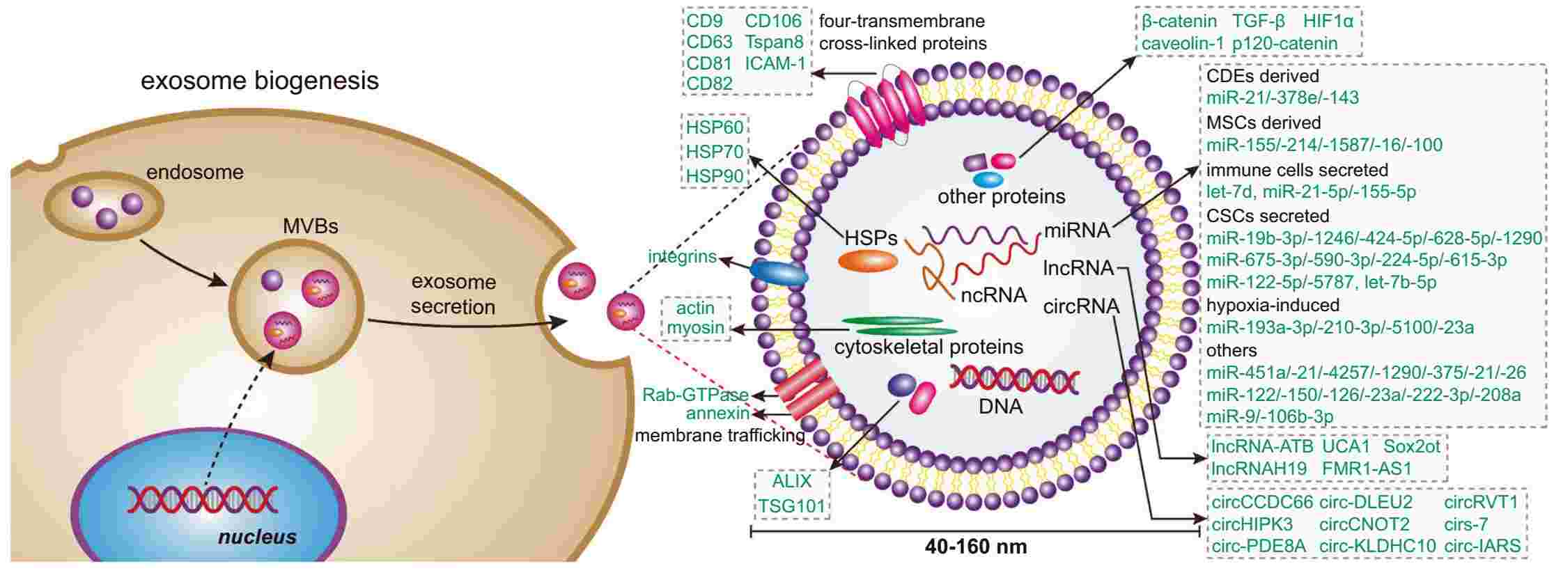 Fig.1 Diagram depicting exosome formation and constituent components.1
How Creative Biolabs' Service Can Assist Your Project?
Creative Biolabs provides an end-to-end analytical suite that transforms PDCD6IP from a mere marker into a dynamic, actionable research component. We deliver not just data, but contextual interpretation, clarifying whether PDCD6IP expression correlates with disease state, identifying its binding partners in pathological conditions, and assessing its utility as a specific exosome tracer. Our solutions directly address prevalent R&D gaps, such as standardizing PDCD6IP measurement across sample types and deconvoluting its role in mixed EV populations.
Uncover the Full Scope of Our Analysis – Initiate a Project Consultation!
Service Workflow
01Project Initiation & Client Input
- Client provides: Purified EVs / Cell Lysates / Biofluids.
- Specific Research Hypotheses (e.g., disease correlation, drug effect).
- Relevant Biological Controls.
02Assay Design & Optimization
Tailoring protocols to specific sample matrices and research goals.
03Sample Processing & Core Execution
- EV Isolation & Characterization.
- PDCD6IP Quantification (MSD/ELISA, Western Blot).
- Interaction & Localization Studies (IP, IF/Confocal).
04Multi-Platform Data Acquisition
Gathering high-fidelity raw data across selected analytical tools.
05Data Integration & Bioinformatics
Correlating molecular data with broader biological contexts.
06Final Reporting & Deliverables
- Comprehensive Technical Report with raw & analyzed data.
- Expert Interpretation & Strategic Recommendations.
- Publication-Ready Figures.
Applications
Our PDCD6IP analysis services are engineered to support a wide spectrum of cutting-edge research and development objectives.
Biomarker Discovery & Validation
Mechanistic Studies in Exosome Biogenesis
Investigate the functional role of PDCD6IP in ESCRT-independent vesicle formation, cargo sorting (e.g., miRNAs, tetraspanins), and its interplay with syntenin-syndecan complexes.
Therapeutic Target Assessment
Evaluate the modulation of PDCD6IP expression or function in response to drug candidates, gene therapies, or RNAi, providing critical MoA (Mechanism of Action) data.
Viral Pathogenesis Research
Study the hijacking of the PDCD6IP/ALIX pathway by viruses for budding, offering insights into novel antiviral strategies.
EV Source & Purity Characterization
Utilize PDCD6IP as a specific exosomal marker to assess the purity of EV preparations and distinguish exosome subpopulations from other extracellular vesicles.
Published Data
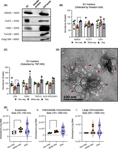 Fig.2 Analysis results used in EV characterizations.2
A pivotal study published employed integrated PDCD6IP analysis to investigate exosome involvement in intervertebral disc degeneration. Researchers isolated sEVs from patient cells and performed rigorous characterization. Quantitative analysis revealed a marked increase in PDCD6IP levels within sEVs from degenerative cohorts compared to controls. This work not only established PDCD6IP as a sensitive biomarker for disc pathology but also suggested its functional role in mediating pathogenic cell-cell communication, highlighting the direct utility of targeted PDCD6IP analysis in defining disease mechanisms and identifying therapeutic avenues.
Why Choose Us?
- Orthogonal Validation Framework: We cross-verify key findings using complementary platforms (e.g., MSD quantification + Western Blot validation + imaging), ensuring unparalleled data reliability and reproducibility.
- Expertise in Complex Matrices: Our optimized protocols are specifically designed for challenging samples like human serum, cerebrospinal fluid (CSF), and conditioned cell media, minimizing artifacts.
- Integrated Data Analysis: We go beyond raw numbers. Our bioinformatics team correlates PDCD6IP data with client-provided omics datasets or our in-house EV characterization data (NTA, TEM).
- Proprietary Assay Development: For unique project needs, our scientists can develop and validate custom immunoassays or imaging protocols targeting specific PDCD6IP isoforms or interaction domains.
Harness the Creative Biolabs Advantage – Connect with Us for a Personalized Quote Today.
Frequently Asked Questions
-
Q: Why is PDCD6IP a better exosome marker than the commonly used CD63 or TSG101?
A: While CD63 and TSG101 are valuable, PDCD6IP/ALIX is a core mechanistic component of the ESCRT-independent biogenesis pathway. Its analysis provides direct functional insight into a specific exosome subpopulation.
-
Q: Can you analyze PDCD6IP in archived tissue samples or formalin-fixed paraffin-embedded (FFPE) sections?
A: Yes. We have optimized immunofluorescence and immunohistochemistry protocols for FFPE tissues, allowing for retrospective analysis of PDCD6IP localization and expression in biobank samples.
-
Q: What sample volume and EV concentration are required for a comprehensive analysis?
A: Requirements vary. For standard quantitative analysis from biofluids, we typically recommend starting with 1-2 mL of serum/plasma or 10-20 mL of conditioned media.
-
Q: Do you provide isolation services for exosomes prior to PDCD6IP analysis?
A: Absolutely. We offer a full suite of EV isolation and characterization services (ultracentrifugation, size-exclusion chromatography) and can seamlessly integrate PDCD6IP analysis as a downstream application.
-
Q: How can PDCD6IP analysis support my IND (Investigational New Drug) application for an EV-based therapy?
A: Our service provides critical Critical Quality Attribute (CQA) data, demonstrating batch-to-batch consistency and confirming the intended cellular source and mechanism for regulatory dossiers.
Our popular analysis services targeting PDCD6IP include but not limited to the following:
| Cat | Service |
| BAS94-1 | PDCD6IP Protein Analysis |
| BAS94-2 | PDCD6IP PTM Analysis Service |
| BAS94-3 | PDCD6IP-XXX interaction Analysis |
| BAS94-4 | Recombinant PDCD6IP Constructs Generation/Characterization |
| BAS94-5 | In Vitro Ubiquitination of PDCD6IP |
| BAS94-6 | PDCD6IP mRNA Analysis |
Contact Us
Creative Biolabs stands as your premier scientific partner for PDCD6IP Analysis Services. For detailed technical specifications, project-specific quotations, or to consult with our senior scientists, please reach out via our website contact form. We are ready to collaborate on your next breakthrough.
References
- Dai, Jie, et al. "Exosomes: key players in cancer and potential therapeutic strategy." Signal transduction and targeted therapy 5.1 (2020): 145. Distributed under CC BY-SA 4.0, without modification. Doi: https://doi.org/10.1038/s41392-020-00261-0.
- Li, Li, et al. "The proteomic landscape of extracellular vesicles derived from human intervertebral disc cells." JOR spine 7.4 (2024): e70007. Distributed under CC BY 4.0, without modification. Doi:https://doi.org/10.1002/jsp2.70007.
For Research Use Only.