We utilize proprietary high-affinity antibodies specifically engineered to recognize various AKAP13 isoforms and post-translational modifications, ensuring minimal cross-reactivity in complex matrices.
Are you currently facing challenges in quantifying scaffold proteins or deciphering complex Rho-guanine nucleotide exchange factor (GEF) signaling pathways in oncology or cardiology? Our AKAP13 Analysis Services help you achieve sensitive, high-throughput detection and functional validation of this multi-domain protein through our advanced immuno-analytical platforms and specialized antibody engineering techniques. Creative Biolabs advances your investigation by optimizing the transition from crude specimens to practical clinical conclusions.
Contact our team to get an inquiry now!
Introduction of AKAP13 Analysis
AKAP13 (also known as BRX, LBC, or Proto-Lbc) is a massive scaffold protein that plays a pivotal role in coordinating spatio-temporal intracellular signaling. As a member of the A-kinase anchoring protein family, its primary structure contains a Dbl homology (DH) domain and a Pleckstrin homology (PH) domain, which collectively function as a Guanine Nucleotide Exchange Factor (GEF) specifically for RhoA. This dual functionality allows AKAP13 to bridge Protein Kinase A (PKA) signaling with the activation of Rho-GTPases, regulating cytoskeletal dynamics, gene transcription via the NF-κB pathway, and steroid hormone receptor sensitization.
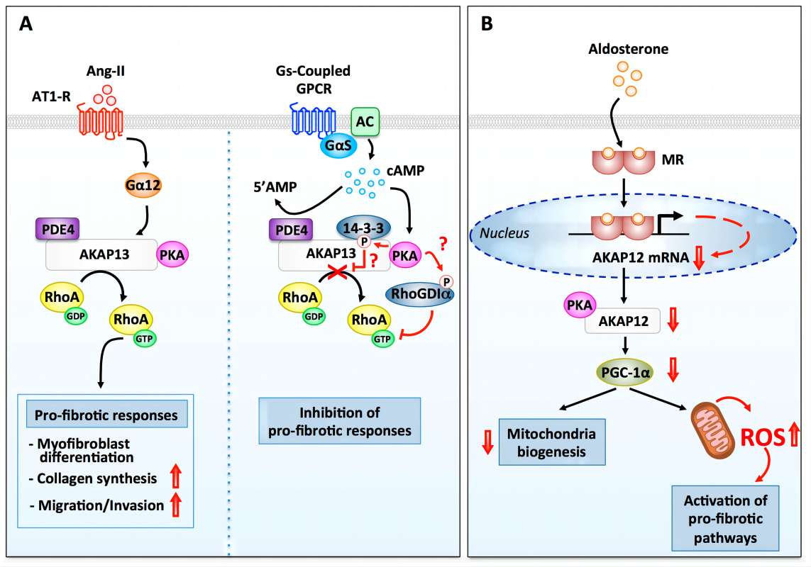 Fig.1 Fibrotic response modulation by AKAP13 in ventricular fibroblasts.1
Extensive literature analysis indicates that AKAP13 is a critical regulator in various diseases. In oncology, its overexpression is linked to the progression of breast, gastric, and prostate cancers, often facilitating epithelial-mesenchymal transition (EMT) and metastasis. Furthermore, AKAP13 is essential for cardiac development and hypertrophic responses, where it mediates the stress-induced activation of p38 MAPK pathways. Given its involvement in complex signaling nodes, precise analysis of AKAP13 expression and its phosphorylation status is indispensable for identifying novel therapeutic targets and developing diagnostic biomarkers for chronic inflammatory and malignant conditions.
Application
Creative Biolabs' AKAP13 analysis spans multiple research and clinical dimensions:
- Oncology Research: Quantitative assessment of AKAP13 as a prognostic biomarker for solid tumors and investigating its role in RhoA-mediated cell migration.
- Cardiovascular Studies: Monitoring AKAP13-dependent signaling in cardiomyocytes to understand the molecular mechanisms behind cardiac hypertrophy and heart failure.
- Drug Target Discovery: High-throughput screening of small molecules or biologics that disrupt the AKAP13-RhoA interaction or inhibit its GEF activity.
- Immune Response Profiling: Analyzing AKAP13's contribution to NF-κB signaling and its impact on chronic inflammatory diseases and innate immunity.
- Diagnostic Assay Development: Utilizing AKAP13 as a component in multiplexed diagnostic panels for early cancer detection and patient stratification.
Service Highlights
Partnering with Creative Biolabs for your AKAP13 evaluation guarantees the utilization of premier scientific proficiency alongside a comprehensive quality control system.
Unrivaled Specificity and Sensitivity
Published Data Reliability
Our platforms are validated against rigorous benchmarks, consistently delivering results that align with peer-reviewed "Published Data" regarding AKAP13's molecular weight and subcellular localization patterns.
Customized Immunoassay Platforms
Whether your project requires Western Blotting, IHC, or ultrasensitive ELISA-based singleplex analysis, we tailor the detection threshold to suit your sample type (serum, tissue, or cell lysates).
Comprehensive Molecular Characterization
Beyond simple quantification, we provide detailed insights into AKAP13 protein-protein interactions (PPIs) using advanced co-immunoprecipitation and mass spectrometry techniques.
Stringent Quality Control
Every project undergoes 100% verification through SDS-PAGE, HPLC, and stability testing, ensuring that the data you receive is ready for regulatory submission or high-impact publication.
Service Workflow
01Initial Consultation and Experimental Design
Our scientists collaborate with you to define the scope, selecting the most appropriate AKAP13-specific reagents and detection platforms based on your biomarker's expected abundance.
02Sample Processing and Preparation
We perform standardized protein extraction and fractionation. For AKAP13, which is often membrane-associated or cytoskeletal, we use specialized lysis buffers to ensure maximum recovery of the target scaffold protein.
03Assay Execution and Optimization
Using Creative Biolabs' optimized protocols, we conduct the primary analysis (e.g., singleplex immunoassay). We perform "fit-for-purpose" validation, adjusting antibody concentrations and incubation times to maximize the signal-to-noise ratio.
04Advanced Data Acquisition
Samples are processed using state-of-the-art imaging and detection systems. We capture raw luminescence, fluorescence, or absorbance data, ensuring linear range accuracy for quantitative comparisons.
05Bioinformatics and Expert Reporting
Data is interpreted by our senior biology specialists. You receive a comprehensive report including raw data, normalized results, statistical significance analysis, and expert commentary on the biological implications of the findings.
FAQs
-
What structural features of AKAP13 complicate its detection via standard immunoassay methods?
AKAP13 is a high-molecular-weight scaffold protein (approximately 300 kDa) containing multiple interaction domains, including DH, PH, and PKA-binding regions. Its tendency to form large protein-protein interaction complexes can lead to epitope masking. Effective quantification requires antibodies with high affinity for accessible surface loops or specific functional domains to overcome steric hindrance and ensure high signal-to-noise ratios.
-
How do the various splice variants of AKAP13 influence analytical specificity?
The AKAP13 gene undergoes extensive alternative splicing, producing variants such as p115-Lbc and BRX which differ in their domain architecture and tissue expression patterns. Specificity is achieved by utilizing antibodies that target unique exon junctions or by employing mass spectrometry-based proteomic workflows to identify signature peptides unique to each isoform, allowing for precise differentiation in heterogeneous samples.
-
What analytical sensitivity is required to monitor AKAP13 in clinical cohorts?
AKAP13 expression is often tightly regulated and may be present at low concentrations in physiological fluids compared to intracellular levels. To detect subtle dysregulation in serum or plasma, analytical platforms must achieve sensitivity in the low picogram per milliliter (pg/mL) range. This level of sensitivity is critical for identifying early-stage compensatory mechanisms in cardiac hypertrophy or metastatic shifts in oncological models.
-
Which pre-analytical factors are most critical for preserving AKAP13 integrity in biological samples?
As a signaling scaffold, AKAP13 is highly susceptible to proteolytic cleavage and phosphorylation changes upon cell lysis. To maintain the native state and concentration of the protein, it is imperative to use rapid cold-chain processing and supplement lysis buffers with a comprehensive cocktail of protease and phosphatase inhibitors. Standardized sample handling is vital to prevent the degradation of the large polypeptide chain.
-
What is the scientific rationale for analyzing AKAP13 levels versus downstream RhoA activation?
While active RhoA levels indicate the immediate state of cellular tension or movement, AKAP13 analysis identifies the specific upstream GEF responsible for integrating multiple inputs (e.g., GPCR and PKA signaling). Measuring AKAP13 provides a more stable biomarker of chronic pathway activation and offers insights into the spatial organization of the signaling nexus, which is often more indicative of long-term disease progression than transient RhoA activity.
Creative Biolabs provides a complete suite of AKAP13 Analysis Services, ranging from custom antibody generation and assay development to high-sensitivity biomarker quantification. Our platform combines deep biological expertise with rigorous QC to accelerate your diagnostic and therapeutic discovery programs.
Reference
- Delaunay, Marion et al. "The Role of Cyclic AMP Signaling in Cardiac Fibrosis." Cells vol. 9,1 69. 26 Dec. 2019, Distributed under Open Access license CC BY 4.0, without modification. https://doi.org/10.3390/cells9010069
For Research Use Only.