We utilize internally developed antibodies that target unique epitopes in the linker region of AKT2, ensuring zero cross-reactivity with AKT1 or AKT3.
Are you currently facing challenges in deciphering complex kinase signaling pathways or struggling with the high background noise in isoform-specific AKT detection during drug screening? Our AKT2 Analysis Services help you achieve definitive isoform-specific quantification and functional validation through Creative Biolabs' proprietary high-affinity antibody platforms and advanced proteomic mass spectrometry. We bridge the gap between discovery and clinical validation, ensuring your therapeutic candidates are evaluated with the highest biological accuracy.
Contact our team to get an inquiry now!
Introduction to AKT2: A Critical Node in Disease Pathogenesis
AKT2, also known as Protein Kinase B beta (PKBβ), is a serine/threonine kinase that serves as a central orchestrator in insulin signaling and glucose metabolism. While the AKT family shares significant structural homology, AKT2 is distinctively localized and functionally specialized. Literature analysis indicates that AKT2 is predominantly expressed in insulin-responsive tissues such as adipose, liver, and skeletal muscle. Structurally, it consists of an N-terminal pleckstrin homology (PH) domain, a central catalytic domain, and a C-terminal regulatory tail containing the hydrophobic motif.
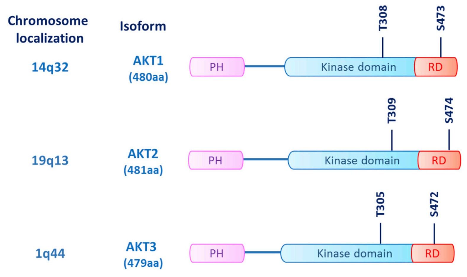 Fig.1 Structural comparison of the three AKT isoforms.1
In the context of oncology, AKT2 is frequently overexpressed or amplified in aggressive phenotypes, particularly in ovarian, pancreatic, and breast cancers. Unlike its sibling AKT1, which is often linked to cell survival, AKT2 is specifically implicated in the epithelial-to-mesenchymal transition (EMT) and the metastatic cascade. Furthermore, dysregulation of the PI3K/AKT2 pathway is a hallmark of Type 2 diabetes and insulin resistance syndromes. Research conclusions emphasize that targeting the unique hydrophobic motif (Ser474) of AKT2 allows for isoform-specific therapeutic intervention, minimizing the systemic toxicity associated with pan-AKT inhibition. Creative Biolabs provides the analytical depth required to navigate these intricate signaling nodes with confidence.
Application of AKT2 Analysis
Creative Biolabs' analysis services are designed to support diverse research and development objectives:
- Isoform-Specific Drug Screening: Distinguishing between AKT1, AKT2, and AKT3 inhibition profiles to identify selective lead compounds with reduced metabolic side effects.
- Biomarker Discovery: Quantifying AKT2 expression levels in patient-derived xenograft (PDX) models or clinical biopsies to correlate with disease progression and treatment response.
- Metabolic Research: Investigating the role of AKT2 in GLUT4 translocation and insulin signaling pathways within adipocytes and myocytes.
- Metastasis Profiling: Analyzing AKT2-mediated phosphorylation of downstream targets like Girdin or Palladin to assess the migratory potential of tumor cells.
- Signaling Pathway Mapping: Utilizing phosphoproteomics to map the crosstalk between AKT2 and other critical pathways such as mTORC1/2 and MAPK.
Service Highlights
Our AKT2 analysis platform stands at the forefront of the industry, offering unmatched specificity and sensitivity.
Proprietary High-Affinity Antibodies
Isoform-Specific Phosphorylation Analysis
Precision detection of phosphorylation at Thr309 and Ser474 using state-of-the-art Multiplex Immunoassays and Western Blotting (Published Data).
Quantitative Mass Spectrometry
Absolute quantification of AKT2 protein levels in complex biological matrices using SRB/MRM-MS techniques.
Functional Validation Assays
Beyond detection, we offer kinase activity assays and siRNA/shRNA-mediated knockdown studies to validate the biological impact of your candidates.
Rigorous Quality Control
Every project is accompanied by a comprehensive technical report including raw data, statistical analysis, and expert interpretation by our senior Ph.D. scientists.
Service Workflow
01Sample Preparation and QC
Total protein extraction using optimized lysis buffers to preserve phosphorylation states. We perform initial quantification to ensure sample integrity meets our strict downstream requirements.
02Isoform-Specific Capture
Utilizing Creative Biolabs' high-selectivity antibodies, we isolate AKT2 through immunoprecipitation or utilize high-sensitivity ELISA plates to partition the isoform from the total AKT pool.
03Data Acquisition
Samples undergo high-resolution detection via capillary electrophoresis (Simple Western) or targeted mass spectrometry. This stage identifies both total protein mass and specific post-translational modifications.
04Bioinformatics & Kinase Activity Profiling
We calculate the ratio of phosphorylated to total AKT2 and integrate these findings with your broader experimental data, such as dose-response curves for inhibitors.
05Comprehensive Final Reporting
Delivery of a detailed analytical dossier, including high-resolution imagery and quantitative data suitable for regulatory filing or high-impact publication.
FAQs
-
How is isoform specificity maintained between AKT2 and AKT1 given their high sequence homology?
The primary challenge in AKT analysis is the 80% sequence identity between isoforms. Specificity is achieved by targeting non-homologous regions, typically within the C-terminal regulatory domain or the hinge region. Methodologies such as competitive peptide displacement and verification using AKT-isoform-deficient cell lines (knockout models) ensure that the analytical signal is derived exclusively from the AKT2 gene product.
-
Which AKT2 phosphorylation sites are most indicative of active signaling?
AKT2 activity is regulated by dual phosphorylation. Initial activation occurs at Thr309 in the activation loop by PDK1, followed by full activation via phosphorylation at Ser474 in the C-terminal hydrophobic motif by mTORC2. Quantitative assessment of the Ser474/Thr309 ratio is a standard metric for evaluating the potentiation of the PI3K pathway.
-
What are the primary considerations when analyzing AKT2 in clinical FFPE specimens?
Formalin fixation can mask epitopes and promote protein cross-linking, which complicates kinase detection. Successful analysis requires optimized heat-induced epitope retrieval (HIER) using high-pH buffers to expose the target residues. Furthermore, because AKT2 phosphorylation is highly transient, the use of phosphatase inhibitors during tissue processing and rapid cold-chain handling are critical for maintaining the biological state of the sample.
-
Can mass spectrometry distinguish between AKT2 mutations and wild-type protein in complex lysates?
Yes. Liquid Chromatography-Tandem Mass Spectrometry (LC-MS/MS) utilizing Multiple Reaction Monitoring (MRM) can differentiate wild-type AKT2 from oncogenic variants, such as the E17K mutation in the PH domain. By monitoring specific transition ions unique to the mutated peptide sequence, absolute quantification of the mutant versus wild-type ratio can be achieved within a single sample.
-
Why is AKT2 specifically targeted in metabolic studies over AKT1 or AKT3?
Genetic ablation studies have demonstrated that AKT2 is the primary isoform responsible for regulating glucose homeostasis. While AKT1 is ubiquitously expressed and linked to growth, AKT2 is the essential mediator of insulin-stimulated GLUT4 translocation to the plasma membrane. Consequently, analyzing AKT2-specific activity is necessary to understand the molecular basis of insulin resistance and Type 2 diabetes.
Creative Biolabs provides industry-leading AKT2 Analysis Services, combining proprietary antibody technology with expert scientific consultation. Whether you are validating a novel therapeutic or exploring basic signaling mechanisms, our platform delivers the precision required for high-stakes biopharmaceutical research.
Reference
- Martorana, Federica et al. "AKT Inhibitors: New Weapons in the Fight Against Breast Cancer?." Frontiers in pharmacology vol. 12 662232. 29 Apr. 2021, Distributed under Open Access license CC BY 4.0, without modification. https://doi.org/10.3389/fphar.2021.662232
For Research Use Only.