Quantitative measurement of serum/plasma AMH levels using high-sensitivity ELISA or CLIA platforms, featuring optimized pre-dilution steps to eliminate C1q-mediated complement interference for superior accuracy.
Creative Biolabs provides comprehensive AMH analysis services, offering multiple custom strategy options to help clients quantify ovarian reserve and investigate reproductive disorders. Our robust protein and gene-level analyses deliver high-precision data for domestic animal research and global human fertility studies.
Introduction of AMH
Anti-Müllerian Hormone (AMH) is a dimeric glycoprotein consisting of two identical subunits linked by sulfide bridges. As a member of the TGF-β superfamily, it undergoes proteolytic cleavage to form a noncovalent complex of N-terminal prodomain (110-kDa) and C-terminal (25-kDa) homodimers, which binds to AMH Receptor II to initiate signaling in the gonads. Secreted exclusively by granulosa cells of pre-antral and small antral follicles, AMH serves as a stable, non-cyclical marker of the functional ovarian reserve pool. Accurate measurement in serum or plasma typically utilizes quantitative sandwich-ELISAs targeting stable epitopes within the pro-region and mature C-terminal domain for maximum clinical reliability.
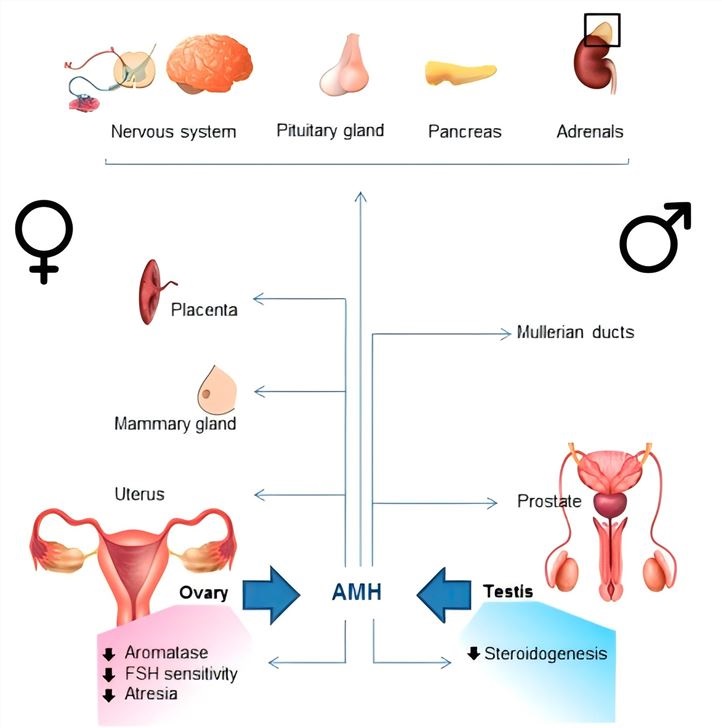 Fig.1 Effects of AMH in the gonads.1
AMH Analysis Services at Creative Biolabs
Creative Biolabs addresses the technical variations in standard automated assays, such as complement interference, by providing a full suite of custom services ranging from core material production to advanced diagnostic development. We prioritize technical performance by validating essential components, including the limit of blank (LOB), linearity, limit of detection (LOD), and limit of quantitation (LOQ) for every custom assay.
Collectively, our featured AMH services include but are not limited to the following:
Hormonal Assay
AMH Polymorphisms Analysis
Genetic screening of variants such as G146T/rs10407022 in the AMH gene and C1749G/rs2071558 in the AMHR2 gene to determine their influence on embryo production and follicle maturation.
Integrated AMH Analysis
A comprehensive multi-omic approach combining protein quantification with genetic profiling to provide a holistic view of the reproductive landscape and hormonal receptor sensitivity.
AMH Antibody Development
Custom generation of high-affinity antibody pairs and antigens validated through IHC and FACS on AMH-positive/negative tissues, supporting applications in sandwich ELISA, LFIA, and TINIA.
Service Workflow
The AMH Analysis workflow is structured to ensure technical precision and data integrity through the following steps:
01Initial Consultation
Clients discuss project objectives and biological targets with our experts to determine the optimal analysis strategy (protein, gene, or integrated).
02Sample Provision
Clients provide initial materials such as serum/plasma for hormonal assays or whole blood/genomic DNA for AMH polymorphism analysis.
03Sample Validation & Pre-treatment
Our laboratory verifies sample quality and performs interference-removal steps, such as specialized pre-dilution to eliminate C1q-mediated complement interference.
04Core Analysis Execution
We perform high-sensitivity quantification via ELISA/CLIA or execute genetic genotyping using validated protocols focused on AMH and AMHR2 variants.
05Quality Control
Every AMH assay is validated against standard performance metrics, including linearity, LOB, LOD, and LOQ, to ensure consistent results.
06Final Delivery
We provide a comprehensive technical report containing raw data, standard curves, and detailed scientific interpretations to guide downstream research or clinical applications.
Applications
Controlled Ovarian Stimulation (COS)
AMH levels serve as a primary predictor for the response to gonadotropins during superovulation protocols. Monitoring this marker allows for precise dosing of medications prior to in vitro fertilization (IVF), significantly reducing the incidence of Ovarian Hyperstimulation Syndrome (OHSS) in high-responders.
Assessment of Ovarian Reserve
Serum AMH provides a reliable quantitative measure of the primordial follicle pool. Because of its strong genomic heritability, it acts as a robust reproductive biomarker for evaluating functional ovarian reserve in patients experiencing infertility or those planning for future family expansion.
Oncofertility and Cytotoxic Impact
Measuring AMH levels is essential for determining the degree of damage to the ovarian reserve caused by chemotherapy or radiotherapy. This analysis helps oncologists and fertility specialists evaluate the gonadotoxic impact of cancer treatments and the recovery potential of ovarian function.
Menopause Transition Prediction
A decline in AMH concentrations often precedes the final menstrual period by several years. Analysis of these levels allows for the prediction of proximity to menopause, assisting women in making informed decisions regarding their reproductive window and hormone replacement therapies.
Diagnosis of Ovarian Disorders
Elevated AMH concentrations are a hallmark of polycystic ovary syndrome (PCOS), reflecting a high volume of small follicles. The analysis provides a biochemical surrogate for antral follicle counts, aiding in the diagnosis of ovulatory dysfunctions and granulosa cell tumors.
Genetic Influence on Embryo Production
Polymorphism analysis in the AMH and AMHR2 genes identifies genetic variations associated with the number of embryos produced during ART. Understanding these variations helps explain discrepancies between hormonal levels and actual clinical outcomes in specific patient populations.
 Fig.2 Applications of AMH detection.
Service Highlights
- Comprehensive Feasibility Study: Every project undergoes an extensive preliminary investigation to evaluate technical potential and resource requirements, ensuring a high prospect for effective solutions and informed decision-making.
- Advanced Interference Management: Our platforms specifically address sample stability issues and complement interference, utilizing optimized pre-dilution protocols to ensure results are accurate and reproducible across different laboratory settings.
- Precise Technical Validation: We provide rigorous validation of LOB, LOD, and LOQ for all assays, delivering industry-leading sensitivity (down to 0.01 ng/mL) and minimal inter-assay variation.
- Expert Project Management: Highly trained professional managers oversee the service from initialization to final closure, ensuring that complex multi-step analyses are delivered on time and according to client specifications.
- Custom Multi-Omic Solutions: By offering integrated analysis of both protein levels and genetic polymorphisms, we provide a deeper level of insight into folliculogenesis than standard diagnostic labs.
FAQs
-
Q: How does Creative Biolabs handle the common issue of complement interference in AMH assays?
A: We implement a specialized sample pre-dilution protocol prior to incubation with capture antibodies. This prevents the binding of C1q and subsequent C3 recruitment, which can otherwise cause falsely low AMH readings in freshly frozen or freshly drawn samples.
-
Q: What is the significance of analyzing AMHR2 polymorphisms?
A: Genetic variations like the C1749G allele in the receptor gene can affect how a patient's ovaries respond to the hormone. Patients with specific genotypes may show poor embryo production even with "normal" serum AMH levels, making this analysis vital for personalized IVF.
-
Q: Can your AMH analysis service be used for oncology patients?
A: Yes, it is the most sensitive method for monitoring the gonadotoxic effects of chemotherapy. We recommend baseline testing before treatment and follow-up analysis post-treatment to assess the degree of ovarian recovery or the onset of premature menopause.
-
Q: Are your reference ranges adjusted for different patient demographics?
A: We provide ethnicity-specific and age-validated reference ranges. This is critical because certain populations, such as East Asian women, may exhibit different diagnostic thresholds for conditions like PCOS compared to European cohorts.
-
Q: How do you validate the specificity of your custom AMH antibodies?
A: We perform rigorous validation using Western Blot, IHC, and FACS on tissues with known positive and negative expression. This ensures that the antibody pairs used in our sandwich ELISAs do not cross-react with other TGF-β family members.
-
Q: Is it possible to use your services for animal model research?
A: Absolutely. Our platform is equipped to handle AMH analysis for domestic animals and laboratory models, providing the same level of sensitivity and linearity required for high-impact veterinary and reproductive research.
Creative Biolabs provides industry-leading AMH analysis services, combining technical precision with genetic insight to optimize reproductive outcomes. Contact us today to develop custom solutions for your research needs.
Reference
- Di Clemente, Nathalie, Chrystèle Racine, and Rodolfo A. Rey. "Anti-Müllerian hormone and polycystic ovary syndrome in women and its male equivalent." Biomedicines 10.10 (2022): 2506. Distributed under Open Access license CC BY 4.0, without modification. https://doi.org/10.3390/biomedicines10102506.
For Research Use Only.