


Capture dynamic suppression patterns over time rather than relying solely on static snapshots.
Creative Biolabs has a tradition of commitment. To achieve efficient execution and regulatory approval, we offer careful considerations of your program for the development of a cellular or gene therapy product – now and in the future.
EXPLORE MORE HighlightsWe focus on unmet needs and develop novel cellular and gene drugs and solutions that offer significant benefits over existing options.
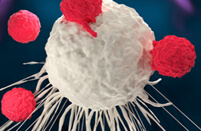
EXPLORE MORE HighlightsThe advent of Chimeric Antigen Receptor T-cell (CAR-T) therapies has revolutionized the field of oncology, providing new avenues for treating various forms of cancer. Our 20 years of experience in the biotechnology sector have equipped us with the expertise and technological capabilities to support the entire lifecycle of CAR-T products, from early development to commercialization.
EXPLORE MORETo accelerate advanced breakthroughs of your projects, we offer broad range of platforms which enable our clients be free to tackle problems with cutting-edge technologies from different angles and in different methods.
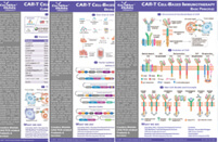
EXPLORE MORE HighlightsUse the resources in our library to help you understand your options and make critical decisions for your study. We offer oncolytic virus, CAR-T, and dendritic cell related documents, as well as newsletter. If you don't find the answers you're looking for, contact us for additional assistance.
EXPLORE MORE HighlightsGet a real taste and understanding of the business and culture of one of the world's great research-based cellular and gene therapy discovery and development companies.
EXPLORE MORE
Understanding the complex, often transient interactions between immune effector cells (IECs) and hematopoietic populations requires more than endpoint measurements. Many suppressive events—such as prolonged contact, serial engagement, or non-lethal immune pressure—occur over time and are not fully captured by static assays. To bridge this gap, live-cell imaging technologies offer a unique advantage: continuous, real-time observation of cellular behavior, motility, and fate decisions within controlled microenvironments.
Creative Biolabs' Live-Cell Imaging based Immune Cell and Blood Cell Interaction Analysis Service is designed to visualize and quantify the dynamic interplay between IECs and hematopoietic targets. As a key module of our ICAHT Management Solutions, this service enables researchers to track contact dynamics, immune activation, target suppression, and recovery trajectories at single-cell and population levels. By integrating cutting-edge imaging platforms with advanced analytical pipelines, we provide mechanistic insights into how immune effectors influence hematopoiesis beyond what traditional assays can reveal.
This service employs real-time imaging strategies—such as widefield microscopy, high-content screening, and time-lapse confocal imaging—to continuously monitor co-cultures of IECs and hematopoietic cells under physiological conditions. We construct custom culture models where CD34⁺ hematopoietic stem/progenitor cells (HSPCs), lineage-committed progenitors, or stromal components interact with CAR-T, CAR-NK, or TCR-engineered cells. These interactions are imaged across multiple time points, often ranging from hours to days, depending on the biological question.
Using fluorescence-labeled immune and target cells, we can non-invasively monitor:
This approach allows researchers to capture how immune cells suppress or eliminate hematopoietic populations in real-time—and under what conditions suppression is reversible, sustained, or avoided altogether.
We utilize state-of-the-art widefield, spinning disk confocal, and high-content imaging systems capable of capturing multiple fluorescence channels simultaneously, with minimal photobleaching and real-time environmental control.
Our system supports 2D, 3D matrix-embedded, and transwell formats for co-culturing immune cells with CD34⁺ HSPCs, stromal cells, or lineage-specific precursors—offering flexibility across various research models.
We apply sophisticated tracking software to monitor individual cell behaviors—including migration, clustering, proliferation, and death—over extended time periods, enabling quantitative kinetic analysis at the single-cell level.
Customized incubation chambers with CO₂, temperature, and humidity regulation allow for continuous, non-disruptive imaging over 72+ hours without affecting cell viability or function.
Our platform allows live detection of key biological processes such as:
Parallel imaging of multiple IEC constructs or conditions (e.g., varying E:T ratios, pre-activation states) allows for robust construct comparison and mechanism-of-action differentiation.
Our scientific team provides fully annotated videos, interaction maps, heatmaps, and quantitative summaries to support data-driven decisions in immune effector optimization or toxicity mitigation.

Capture dynamic suppression patterns over time rather than relying solely on static snapshots.
Directly measure cell engagement frequency, duration, and cytotoxic vs. non-lethal outcomes.
Use 2–4 fluorescent channels simultaneously to track multiple cell types and intracellular events.
Compatible with a wide range of immune effectors (CAR-T, CAR-NK, TCR) and human hematopoietic targets (CD34⁺, MPPs, stromal cells).
Visualize whether hematotoxicity results from prolonged immune contact, motility restriction, swarm behavior, or paracrine cues.
Q1: What if my immune cells do not naturally express fluorescent markers?
A1: We can assist in labeling with non-toxic dyes or transient transduction with fluorescent reporters, ensuring minimal interference with cell function.
Q2: Is this service suitable for suspension cells?
A2: Yes. We use specialized imaging chambers and low-adhesion coatings to maintain optimal positioning of suspension cells for time-lapse microscopy.
Q3: Can I request custom time intervals or imaging durations?
A3: Absolutely. Imaging frequency, duration, and resolution can be adjusted based on your biological question and cell behavior kinetics.
At Creative Biolabs, we understand that understanding the how is just as important as the what. Our Live-Cell Imaging based Immune Cell and Blood Cell Interaction Analysis Service enables researchers to visualize, quantify, and interpret IEC-induced hematopoietic modulation in real time—offering a unique lens into immune-blood cell dynamics.
Let's work together to build safer, mechanism-informed immune effector cell therapies. Connect with our team today to explore how live-cell imaging can accelerate your understanding of immune–hematopoietic crosstalk.
For any technical issues or product/service related questions, please leave your information below. Our team will contact you soon.
All products and services are For Research Use Only and CANNOT be used in the treatment or diagnosis of disease.
 NEWSLETTER
The latest newsletter to introduce the latest breaking information, our site updates, field and other scientific news, important events, and insights from industry leaders
LEARN MORE NEWSLETTER NEW SOLUTION
CellRapeutics™ In Vivo Cell Engineering: One-stop in vivo T/B/NK cell and macrophage engineering services covering vectors construction to function verification.
LEARN MORE SOLUTION NOVEL TECHNOLOGY
Silence™ CAR-T Cell: A novel platform to enhance CAR-T cell immunotherapy by combining RNAi technology to suppress genes that may impede CAR functionality.
LEARN MORE NOVEL TECHNOLOGY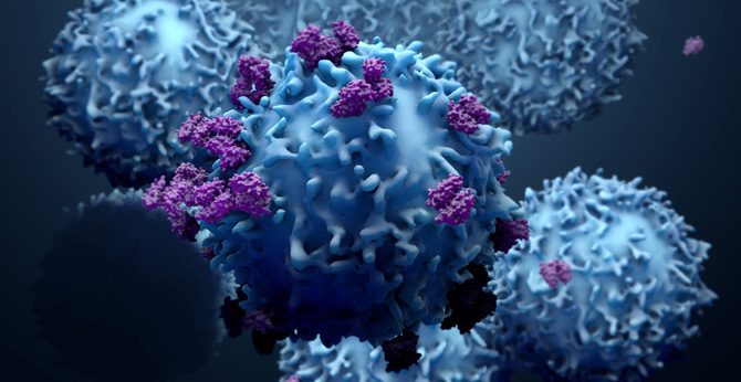 NEW SOLUTION
Canine CAR-T Therapy Development: From early target discovery, CAR design and construction, cell culture, and transfection, to in vitro and in vivo function validation.
LEARN MORE SOLUTION