Ebola Virus Glycoprotein: Structure, Entry Mechanism, and Therapeutic Implications
The Ebola virus (EBOV) remains a catastrophic global health threat, causing Ebola Virus Disease (EVD) with mortality rates often surpassing 50%. Its ability to cripple host immune defenses and trigger severe systemic damage poses persistent risks, especially in resource-constrained regions. Central to EBOV's pathogenicity is its envelope glycoprotein (GP)—a versatile molecular "key" that mediates host cell invasion. Deciphering GP's structure and function is pivotal for unraveling viral infection mechanisms and advancing effective antivirals and vaccines. This article provides a concise analysis of EBOV GP, from its molecular architecture to its role in viral entry, and explores its translational potential in combating EVD.
Ebola Virus GP: Molecular Anatomy
EBOV GP's complexity originates from its unique genetic expression. The GP gene undergoes transcriptional editing, producing three primary isoforms: soluble glycoprotein (sGP), implicated in immune modulation; small soluble glycoprotein (ssGP), with uncharacterized functions; and transmembrane full-length GP—the trimeric spike protein critical for viral entry and pathogenesis.
Full-length GP (GP1,2) is a trimer composed of disulfide-linked GP1 (receptor-binding subunit) and GP2 (membrane fusion subunit). GP1 features a receptor-binding domain (RBD), a glycosylated glycan cap, and a mucin-like domain (MLD) densely decorated with O-linked and N-linked glycans. GP2 contains a hydrophobic fusion loop (FL), heptad repeat regions (HR1, HR2), a transmembrane domain (TM), and a cytoplasmic tail (CT).
X-ray crystallography and cryo-electron microscopy (cryo-EM) have uncovered GP's 3D structure, revealing pre-fusion and post-fusion conformations. The pre-fusion trimer is stabilized by intersubunit interactions, while the post-fusion state forms a stable six-helix bundle (6-HB) via HR1-HR2 interactions— a signature of class I viral fusion proteins. Glycosylation plays dual roles: it acts as a "molecular camouflage" to evade antibodies and modulates protein folding, stability, and interactions with host lectins like DC-SIGN.
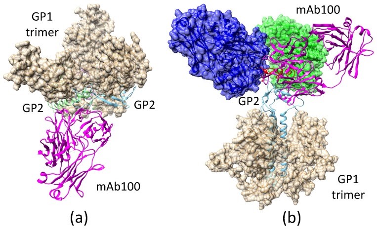 Fig.1 Binding of mAb100 to EBOV GP trimer in pre-fusion and fusion-initiation states.1,2
Dynamic Invasion: GP-Mediated Viral Entry
EBOV's cell invasion is a coordinated process driven by GP, encompassing five core steps:
- Attachment and Receptor Recognition
EBOV attaches to host cells via GP interactions with surface molecules such as C-type lectins (DC-SIGN/L-SIGN), TIM/TAM family proteins, and integrins. These bind to GP's glycosylated MLD or RBD, with receptor redundancy enabling broad cell tropism.
- Endocytic Internalization
Following attachment, the virus enters cells primarily via clathrin-mediated endocytosis (or macropinocytosis). GP's interaction with host proteins triggers endocytic vesicle formation, encapsulating the virus for intracellular transport.
- Endosomal Trafficking and Low-pH Triggering
Virus-containing endosomes mature from early to late stages, where the acidic lumen (pH 5.0–5.5) induces GP's irreversible conformational rearrangement—essential for fusion.
- Membrane Fusion
Low pH destabilizes the pre-fusion trimer, causing GP1 dissociation and exposing GP2's fusion loop. The loop inserts into the endosomal membrane, and HR1-HR2 rearrangement forms a 6-HB, generating force to fuse viral and host membranes.
- Genome Release
A fusion pore expands, releasing the viral nucleocapsid into the cytoplasm to initiate replication. Key residues (e.g., pH-sensing histidines, hydrophobic fusion loop residues) link GP's structure to its function.
Services you may interested in
GP and the Immune System: Evasion vs. Neutralization
GP is central to the "arms race" between EBOV and the host immune system. Its MLD and glycan cap's dense glycosylation shields neutralizing epitopes, while secreted sGP acts as an antibody "decoy," diverting immune responses. GP may also interfere with innate immune pathways (e.g., interferon signaling) via its cytoplasmic tail.
Despite evasion tactics, host neutralizing antibodies target conserved, glycan-free epitopes—such as the GP trimer base or GP1-GP2 interface. Structural biology has guided the identification of these epitopes, enabling optimized antigen design for vaccines and therapies.
Translational Applications: GP-Based Interventions
GP's critical roles make it a prime target for EVD prevention and treatment:
- Vaccines
GP is the core antigen for viral vector, recombinant protein, and mRNA/DNA vaccines. Structural insights have led to optimized variants (e.g., MLD-deficient GPΔmuc, pre-fusion-stabilized GP) that expose more neutralizing epitopes, enhancing immune responses.
- Antivirals
GP-targeting antivirals include entry inhibitors (blocking GP-receptor binding) and fusion inhibitors (disrupting 6-HB formation, e.g., HR2-derived peptides). Neutralizing antibody cocktails, targeting conserved GP epitopes, also show promise.
- Diagnostics
GP's specificity enables serological tests (e.g., ELISAs) to detect anti-GP antibodies and rapid lateral flow assays to identify viral proteins, supporting early outbreak detection.
2024–2025 Research Advances and Future Directions
Recent breakthroughs have deepened GP understanding:
- Structural Technology: High-resolution cryo-EM and cryo-electron tomography (cryo-ET) have visualized GP's dynamic intermediate states and native viral surface distribution.
- Novel Host Interactions: New host proteins and lipids regulating GP-mediated entry and replication have been identified, offering alternative therapeutic targets.
- Non-Canonical Functions: GP's cytoplasmic tail and sGP may regulate viral budding, cell-to-cell spread, and host signaling (e.g., apoptosis, inflammation).
Future challenges include developing universal vaccines/therapies against EBOV species and filoviruses, overcoming GP genetic variability, and studying GP function in live models. Integrating structural data with system-level host immune analyses will further advance EVD intervention design.
In summary, EBOV GP is a multifunctional molecular machine central to viral pathogenesis. Decades of research have translated structural and functional insights into promising interventions. As technology and interdisciplinary collaboration progress, we move closer to controlling EVD—a threat that once seemed insurmountable.
If you want to learn more about the norovirus vaccine, please refer to:
Designing mRNA Ebola Vaccines Delivery Systems and Immunogenicity Analysis
Ebola Virus Replication Cycle Host Factors-RdRp Kinetics-and Vaccine Targets
Browse our Norovirus Antigen Products
→ Ebola Virus - Viral Antigens
Need a custom solution? If our off-the-shelf products aren't a perfect fit, we can create one for you. Contact us to design a product that precisely matches your experimental demands.
References
- Lappala, Anna, et al. "Structural transition and antibody binding of EBOV GP and ZIKV E proteins from pre-fusion to fusion-initiation state." Biomolecules 8.2 (2018): 25. https://doi.org/10.3390/biom8020025
- Distributed under Open Access license CC BY 4.0, without modification.
Created December 2025
All of our products can only be used for research purposes. These vaccine ingredients CANNOT be used directly on humans or animals.
