The Molecular Architecture of HPV: Decoding the L1 Capsid Protein
Introduction: Why the L1 Protein Is Key to Understanding HPV
Human papillomavirus (HPV) represents a significant global public health challenge, associated with a range of diseases from benign warts to various cancers. Understanding the structural components of the virus is essential for developing effective preventive strategies. At the heart of the viral structure lies the capsid, a protective shell that encloses the viral genome and facilitates infection. The L1 capsid protein is the fundamental building block of this shell, serving as the major viral capsid protein in HPV. This article delves into the molecular structure, immunological properties, and vaccine applications of the L1 protein, highlighting its role as the cornerstone of modern HPV prevention.
Basic Biology of the L1 Protein: The Blueprint of HPV Virion Assembly
Definition and Function: What Is the HPV L1 Capsid Protein?
The L1 protein is the primary structural component of HPV, accounting for 80–90% of the total viral capsid mass. Its main function is to self-assemble into the protective outer shell of the virus, known as the capsid. Through a highly efficient process, L1 monomers form pentamers, also called capsomeres. Seventy-two of these pentameric units then assemble into a complete icosahedral symmetric structure, forming the mature virion.
The Marvel of Self-Assembly: Virus-Like Particles (VLPs) and Their Significance
One of the most remarkable features of the L1 protein is its ability to spontaneously form virus-like particles (VLPs) in the absence of viral DNA or other viral proteins such as L2. These VLPs closely mimic the size and morphology of native HPV virions but are non-infectious and non-pathogenic since they lack genetic material. Their structural authenticity allows them to elicit strong immune responses, making VLPs ideal antigens for recombinant HPV vaccines.
Service you may interested in
High-Resolution Insights into the L1 Protein: Unveiling Its Three-Dimensional Structure
Cutting-Edge Techniques: How Cryo-EM and X-Ray Crystallography Visualize the L1 Protein
Advanced structural biology techniques, including cryo-electron microscopy (cryo-EM) and X-ray crystallography, have been instrumental in elucidating the architecture of the L1 protein. These methods have overcome challenges such as particle flexibility and heterogeneity to provide atomic-level insights into the capsid organization.
A Milestone in Structural Biology: High-Resolution Structure of the HPV16 L1 Capsid
A landmark study published in 2020 reported the cryo-EM structure of the HPV16 capsid at 3.1 Å resolution. This breakthrough not only provided a detailed atomic model of the L1 capsid protein but also revealed the density of the minor capsid protein L2 and highlighted conformational flexibility within the capsid. The atomic coordinates are available in the Protein Data Bank under accession code 7KZF.
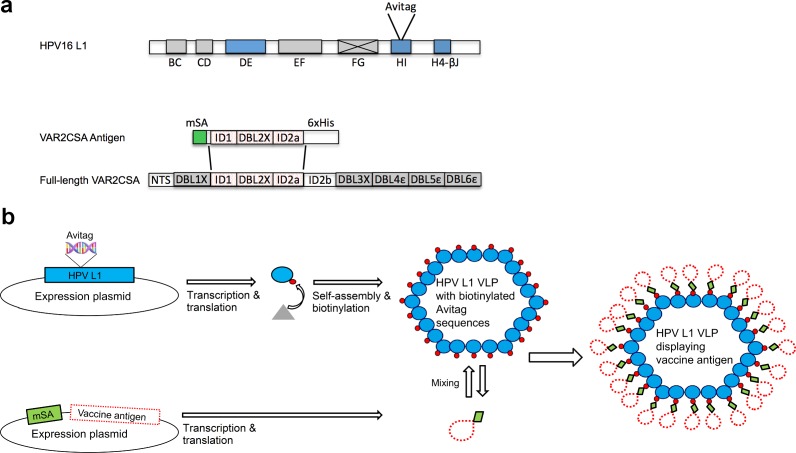 Fig.1 Diagram of VAR2CSA VLP-Vaccine Assembly and Components.1,2
Key Structural Domains and Their Functions: A Parts List of the L1 Protein
The L1 protein adopts a conserved β-jelly roll fold, common among many viral capsid proteins. Exposed surface loops—BC, DE, EF, FG, and HI—exhibit conformational variability and host critical neutralizing epitopes. Additionally, C-terminal and N-terminal arms facilitate inter-pentamer connections, ensuring the stability and integrity of the icosahedral capsid.
Immunological Characteristics of the L1 Protein: A Target for Vaccine Design
Why L1 Is a Potent Immunogen: The Natural Advantage of VLPs
The highly repetitive and ordered surface of VLPs mimics natural viral infection, efficiently presenting antigenic epitopes to immune cells. This results in a robust humoral immune response, forming the basis for the high efficacy of L1-based HPV vaccines.
Decoding Immunogenic Epitopes: Precision Targets for Neutralizing Antibodies
Most neutralizing epitopes on the L1 protein are conformational, meaning they depend on the three-dimensional structure rather than linear amino acid sequences. Key immunogenic regions are located within surface-exposed loops, particularly the FG and HI loops. Although epitope mapping continues, much of the current knowledge relies on well-established data.
Methodologies for Epitope Identification: How Scientists Locate These Targets
Common techniques for epitope characterization include:
- VLP-based ELISA: A standard method for detecting antibody reactivity.
- Pseudovirus-based neutralization assay (PBNA): A functional test to evaluate antibody-mediated neutralization.
- Monoclonal antibody mapping: Uses specific antibodies to pinpoint binding sites.
- Peptide microarray: Enables high-throughput screening of linear epitopes
Core Technology of Modern HPV Vaccines
The VLP Technology Revolution: Why HPV Vaccines Are Subunit Vaccines
All commercially available prophylactic HPV vaccines are subunit vaccines. They are produced by expressing the L1 capsid protein in recombinant systems, allowing it to self-assemble into VLPs, which are then purified and used as antigens. This approach ensures high safety and potent immunogenicity.
An Overview of Mainstream HPV Vaccine Technologies: From Expression Systems to Adjuvant Formulations
All commercially available prophylactic HPV vaccines are subunit vaccines. They are produced by expressing the L1 capsid protein in recombinant systems, allowing it to self-assemble into VLPs, which are then purified and used as antigens. This approach ensures high safety and potent immunogenicity.
Comparison of Major HPV VLP Vaccine Production Platforms
| Feature |
Yeast-Expression Platform (Used for quadrivalent & nonavalent vaccines) |
Baculovirus-Insect Cell Platform (Used for bivalent vaccine) |
| Expression System |
Recombinant Yeast (Saccharomyces cerevisiae) |
Baculovirus Expression Vector System (BEVS) (In Trichoplusia ni insect cells) |
| Purification Process | Multi-step process involving fermentation, cell disruption, and extensive chromatographic and filtration methods for purification before VLP self-assembly. | Multi-step process involving cell culture and purification through various chromatography and filtration steps. |
| Adjuvant Formulation | Aluminum-based adjuvant (Amorphous Aluminum Hydroxyphosphate Sulfate) | Proprietary Adjuvant System combining Aluminum Hydroxide and a TLR4 Agonist (Monophosphoryl lipid A) to enhance immune response. |
Conclusion and Future Perspectives: The Past and Future of the L1 Protein
The L1 capsid protein is fundamental to HPV virion assembly. Its ability to form VLPs has revolutionized vaccine development, leading to highly effective preventive tools against HPV-related diseases. Future research may focus on developing broader-spectrum vaccines, engineering L1 for improved immunogenicity or stability, and exploring therapeutic applications through a deeper understanding of L1-immune system interactions
Ready to advance your Human Papillomavirus (HPV) vaccine project? Creative Biolabs is your expert partner, offering a comprehensive suite of services from innovative vaccine design and construction to rigorous preclinical testing. Our seasoned scientists are dedicated to helping you navigate the complexities of development and achieve your research milestones with greater speed and efficiency. If you have any needs in these areas, don't hesitate to contact our experts. Let's collaborate to take your vital scientific research to the next level.
If you want to learn more about the humanized mice, please refer to:
From Lab to Jab: The Scientific Development Timeline of the HPV Vaccine
Ready-to-Use HPV Antibodies
| CAT | Product Name | Target | Type | Price |
|---|---|---|---|---|
| VAnt-Wyb178 | HPV Monoclonal Antibody (E7, IgG2a, 3.3 mg/ML) | Human Papillomavirus | Antibody | Inquiry |
| VAnt-Wyb179 | HPV Monoclonal Antibody (E7, IgG1, 3.8 mg/ML) | Human Papillomavirus | Antibody | Inquiry |
| VAnt-Wyb180 | HPV Monoclonal Antibody (E7, IgG2a, 3.4 mg/ML) | Human Papillomavirus | Antibody | Inquiry |
| VAnt-Wyb181 | HPV Monoclonal Antibody (E6, IgG1-κ, 4.8 mg/ML) | Human Papillomavirus | Antibody | Inquiry |
| VAnt-Wyb182 | HPV Monoclonal Antibody (E6, IgG1-κ, 1.01 mg/ML) | Human Papillomavirus | Antibody | Inquiry |
| VAnt-Wyb183 | HPV Monoclonal Antibody (L1, IgG1, 3.0 mg/ML) | Human Papillomavirus | Antibody | Inquiry |
| VAnt-Wyb184 | HPV Monoclonal Antibody (L1, IgG2a-κ, 4.5 mg/ML) | Human Papillomavirus | Antibody | Inquiry |
| VAnt-Wyb185 | HPV Monoclonal Antibody (L1, IgG2a-κ, 3.3 mg/ML) | Human Papillomavirus | Antibody | Inquiry |
| VAnt-Wyb186 | HPV Monoclonal Antibody (E7, IgG2b, 4.3 mg/ML) | Human Papillomavirus | Antibody | Inquiry |
| VAnt-Wyb187 | HPV Monoclonal Antibody (E7, IgG2a, 4.4 mg/ML) | Human Papillomavirus | Antibody | Inquiry |
| VAnt-Wyb188 | HPV Monoclonal Antibody (E7, IgG1, 3.0 mg/ML) | Human Papillomavirus | Antibody | Inquiry |
| VAnt-Wyb189 | HPV Monoclonal Antibody (E7, IgG2a, 5.1 mg/ML) | Human Papillomavirus | Antibody | Inquiry |
| VAnt-Wyb190 | HPV Monoclonal Antibody (E7, IgG2a, 4.7 mg/ML) | Human Papillomavirus | Antibody | Inquiry |
| VAnt-Wyb191 | HPV Monoclonal Antibody (Clone: VA-1870H, IgG1-κ, 1 mg/ML) | Human Papillomavirus | Antibody | Inquiry |
| VAnt-Wyb192 | HPV Monoclonal Antibody (Clone: VA-187H, IgG1-κ, 1 mg/ML) | Human Papillomavirus | Antibody | Inquiry |
| VAnt-Wyb193 | HPV Monoclonal Antibody (Clone: VA-187H, IgG1, 7.4 mg/ML) | Antibody | Inquiry |
Browse our HPV Antigen Products
Need a custom solution? If our off-the-shelf products aren't a perfect fit, we can create one for you. Contact us to design a product that precisely matches your experimental demands.
Reference
- Thrane, Susan, et al. "A novel virus-like particle based vaccine platform displaying the placental malaria antigen VAR2CSA." PloS one 10.11 (2015): e0143071. https://doi.org/10.1371/journal.pone.0143071
- Distributed under Open Access license CC BY 4.0, without modification.
All of our products can only be used for research purposes. These vaccine ingredients CANNOT be used directly on humans or animals.
