Creative Biolabs has a tradition of commitment. To achieve efficient execution and regulatory approval, we offer careful considerations of your program for the development of a cellular or gene therapy product – now and in the future.
EXPLORE MORE HighlightsWe focus on unmet needs and develop novel cellular and gene drugs and solutions that offer significant benefits over existing options.
EXPLORE MORE HighlightsThe advent of Chimeric Antigen Receptor T-cell (CAR-T) therapies has revolutionized the field of oncology, providing new avenues for treating various forms of cancer. Our 20 years of experience in the biotechnology sector have equipped us with the expertise and technological capabilities to support the entire lifecycle of CAR-T products, from early development to commercialization.
EXPLORE MORETo accelerate advanced breakthroughs of your projects, we offer broad range of platforms which enable our clients be free to tackle problems with cutting-edge technologies from different angles and in different methods.
EXPLORE MORE HighlightsUse the resources in our library to help you understand your options and make critical decisions for your study. We offer oncolytic virus, CAR-T, and dendritic cell related documents, as well as newsletter. If you don't find the answers you're looking for, contact us for additional assistance.
EXPLORE MORE HighlightsGet a real taste and understanding of the business and culture of one of the world's great research-based cellular and gene therapy discovery and development companies.
EXPLORE MORE
Despite the clinical success of CAR-T therapy, distinguishing functional memory phenotypes from dysfunctional exhausted states within the tumor microenvironment remains a major challenge, limiting the accurate prediction of long-term antitumor responses. To address this, we present Multi-color Flow Cytometry for In vivo CAR-T Phenotypic Profiling: Memory vs. Exhaustion, a specialized analytical approach. Our service provides high-dimensional immunophenotyping of key surface and intracellular markers on CAR-T cells directly from in vivo samples. By simultaneously resolving memory- and exhaustion-associated signatures, we deliver precise, quantitative profiling that empowers you to optimize therapeutic efficacy and assess persistence potential.
CAR-T cells exhibit diverse phenotypic states that fundamentally determine their functional fate and therapeutic efficacy. Among these, memory CAR-T cells are characterized by high proliferative capacity and long-term persistence, whereas exhausted CAR-T cells display progressive loss of effector function and sustained expression of inhibitory receptors. Accurately distinguishing memory from exhaustion phenotypes is therefore critical for predicting antitumor responses, optimizing CAR design, and guiding combination strategies to improve clinical outcomes.
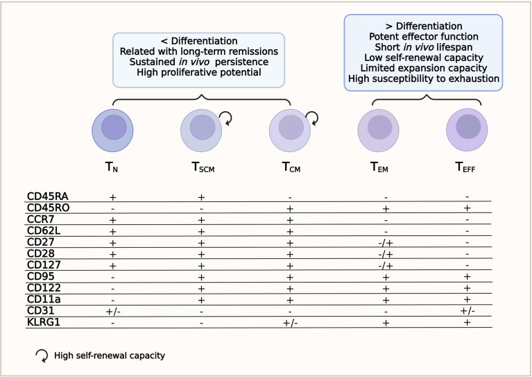 Fig.1 The relevant phenotypic and biological features of distinct T cell subsets that contribute to the success of CAR-T cell therapy.1
Our Multi-color Flow Cytometry service enables simultaneous in vivo profiling of memory and exhaustion markers on CAR-T cells. By resolving key phenotypic signatures, it distinguishes functional from dysfunctional states, providing precise immunophenotyping. This approach supports optimized therapeutic assessment, enhances prediction of antitumor persistence, and empowers preclinical and translational CAR-T research with high-dimensional data quality.
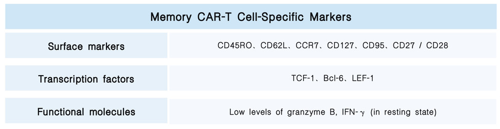
Memory CAR-T cells typically exhibit high proliferative capacity, rapid effector function, and long-term persistence.
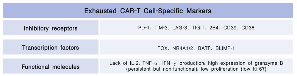
Exhausted CAR-T cells are characterized by progressive loss of effector function, decreased proliferative capacity, sustained high expression of inhibitory receptors, and metabolic abnormalities.
Q1: How is an individual cell suspension prepared from tumor tissue, and is assistance required from your team?
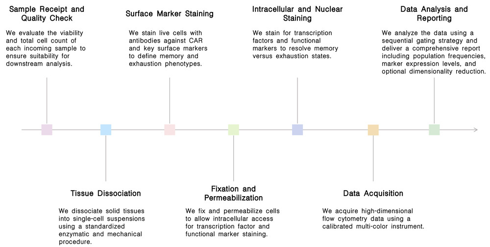
A1: We provide a standardized tissue dissociation protocol utilizing an enzyme cocktail combined with mechanical dissociation. Clients may send fresh or frozen tissue samples to us, and we will perform the complete workflow including dissociation, staining, and flow cytometry acquisition. Alternatively, we can supply a detailed protocol for clients who prefer to prepare the individual cell suspension themselves prior to sample submission.
Q2: How do you distinguish resting memory CAR-T cells from early activated CAR-T cells, given that both typically exhibit low PD-1 and low granzyme B expression?
A2: We utilize CD25 and CD69 as early activation markers to resolve this distinction. Resting memory CAR-T cells are characterized as CD25⁻CD69⁻ with high TCF-1 expression, whereas early activated cells are CD25⁺ and/or CD69⁺, with TCF-1 remaining positive but potentially showing low-level PD-1 upregulation. If required, we can incorporate CD25 and CD69 into our panel to enable this functional discrimination.
We deliver high-dimensional, standardized flow cytometry panels that simultaneously resolve memory and exhaustion signatures in vivo. Our validated protocols, rigorous quality controls, and expert data interpretation ensure reproducible, clinically relevant profiling. Choose us to accurately assess CAR-T functional status and optimize therapeutic strategies with confidence.
"The standardized multi-color panel and rigorous quality controls allowed us to reproducibly track memory versus exhaustion kinetics across multiple mouse models. The inclusion of early activation markers (CD25, CD69) resolved the ambiguous 'low PD-1' populations that previously confounded our data. I highly recommend this service for any translational CAR-T study." Dr. James O'Brien.
"What impressed us most was the ability to simultaneously profile surface, transcription factor, and functional markers from a single tumor biopsy. The team provided clear gating strategies, controls, and a comprehensive report that correlated memory phenotype with in vivo persistence. This service has become an essential quality control step before our product release." Dr. Mei-Lin Chen.
Reach out to our expert team today to discuss your specific CAR-T phenotyping needs. Whether you require a customized panel design, a detailed quote, or technical guidance, we are ready to assist. Simply complete the inquiry form on this page, and we will respond promptly to accelerate your research.
Reference
For any technical issues or product/service related questions, please leave your information below. Our team will contact you soon.
All products and services are For Research Use Only and CANNOT be used in the treatment or diagnosis of disease.
 NEWSLETTER
The latest newsletter to introduce the latest breaking information, our site updates, field and other scientific news, important events, and insights from industry leaders
LEARN MORE NEWSLETTER NEW SOLUTION
CellRapeutics™ In Vivo Cell Engineering: One-stop in vivo T/B/NK cell and macrophage engineering services covering vectors construction to function verification.
LEARN MORE SOLUTION NOVEL TECHNOLOGY
Silence™ CAR-T Cell: A novel platform to enhance CAR-T cell immunotherapy by combining RNAi technology to suppress genes that may impede CAR functionality.
LEARN MORE NOVEL TECHNOLOGY NEW SOLUTION
Canine CAR-T Therapy Development: From early target discovery, CAR design and construction, cell culture, and transfection, to in vitro and in vivo function validation.
LEARN MORE SOLUTION