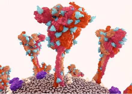
Glycosylphosphatidylinositol Anchor Analysis Service
Glycosylphosphatidylinositol (GPI) anchors serve as lipid moieties that tether proteins to the outer leaflet of the plasma membrane, offering an alternative to transmembrane domains. The structural diversity and functional relevance of GPI-anchored proteins (GPI-APs) span numerous biological processes—including signal transduction, immune modulation, and pathogen-host interactions. At Creative Biolabs, we provide a dedicated GPI anchor analysis service for researchers studying membrane-tethered proteins, post-translational lipid modifications, or GPI biosynthesis-related disorders.
Why GPI Anchors Matter in Biology?
GPI-anchored proteins are synthesized in the endoplasmic reticulum (ER) and undergo post-translational attachment via a conserved glycolipid core. They are localized in lipid rafts, facilitating cell signaling, adhesion, and enzymatic activity. GPI-Aps are vital in various biological processes, including:
- Cell Surface Localization: GPI anchors direct proteins to lipid rafts, influencing membrane trafficking and protein stability.
- Immune Regulation: GPI-anchored proteins serve as complement regulators, co-stimulatory molecules, and adhesion mediators.
- Neural Function: Critical for synaptic development and signal transmission in the nervous system.
- Disease Association: Defects in GPI biosynthesis lead to inherited GPI deficiencies (IGDs) and paroxysmal nocturnal hemoglobinuria (PNH).
GPI Anchor Biosynthesis Pathway
GPI anchors comprise a phosphatidylinositol moiety, a conserved glycan core (GlcN, Man₃), and an ethanolamine phosphate (EtNP) bridge that connects the anchor to the protein's C-terminus. Its biosynthetic pathway includes over 20 genes, grouped into three stages:
1. Early ER Steps
- Transfer of GlcNAc to PI (via PIG-A, PIG-H).
- Deacetylation, acylation, and mannose addition (PIG-M, PIG-V).
2. Protein Attachment
- Mediated by GPI transamidase complex (PIG-K, GPAA1), which replaces the C-terminal signal peptide.
3. Anchor Remodeling
- Fatty acid modifications and EtNP side chain remodeling in the Golgi.
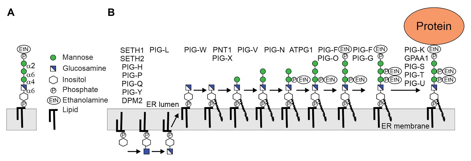 Fig.1 (A) Conserved GPI-anchor backbone structure. (B) Plant GPI precursor biosynthesis in the ER.1
Our GPI Anchor Analysis Workflow
Creative Biolabs offers a comprehensive and customizable workflow to identify, enrich, and characterize GPI-anchored proteins across species and sample types.
- Sample Preparation: Membrane protein extraction under GPI-preserving conditions.
- Enrichment of GPI-Aps: Affinity chromatography.
- Mass Spectrometry (LC-MS/MS): Detection of peptide fragments containing GPI-specific residues.
- Bioinformatic Mapping: Identification of GPI anchor attachment sites and structural variations.
- Quantitative Reporting: Abundance, modification state, and potential biosynthetic defects.
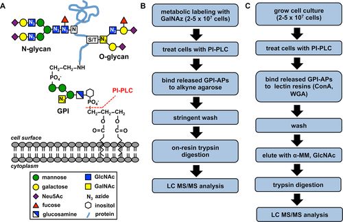 Fig.2 GPI-AP structure and workflows for sugar analog (B) and lectin affinity (C) capture enrichment.2
Frequently Analyzed Targets
| Protein | Application |
|---|---|
| CD55 (DAF), CD59 | Complement regulation, PNH diagnostics |
| PrP (Prion Protein) | Neurodegeneration, infectious disease research |
| Folate receptor (FOLR1) | Cancer targeting, nutrient uptake |
| Thy-1, NCAM | Neurobiology, signal transduction |
| Tetherin (BST2) | Host-virus interaction, innate immunity |
Samples & Deliverables
| Sample Types | Cell lysates, tissue homogenates, purified protein fractions |
|---|---|
| Turnaround Time | Typically, 10–20 business days per analysis cycle |
| Deliverables | Custom report including: methodology, raw data, annotated spectra, GPI peptide mapping, and comparative quantification |
Applications of GPI Anchor Analysis
- Protein localization studies
- Lipid raft composition in immunology or neuroscience
- Paroxysmal Nocturnal Hemoglobinuria (PNH)
- Inherited GPI Deficiency (IGD)
- Prion diseases and GPI-linked neurodegeneration
- Functionalization of recombinant proteins via GPI
- Engineering of GPI-anchored therapeutic enzymes or antigens
Why Choose Creative Biolabs?
- Decades of experience in glycolipid research ensure accurate interpretation of GPI anchor structure and function.
- Advanced LC-MS/MS platforms enable high-sensitivity detection of lipid-linked glycopeptides.
- Expertise in GPI biosynthesis pathways supports defect mapping and anchor remodeling analysis.
- Customized protocols optimize glycolipid analysis across diverse sample types and species.
- Proven support for translational projects involving GPI-anchored proteins in immunity and disease.
Whether you're working on GPI-anchored protein L, studying GPI anchor attachment 1 protein, or reviewing novel GPI-anchored protein line structures, our service is built to adapt to your research scope. Contact us today to request a quote or consult on custom GPI anchor profiling strategies tailored to your project.
References:
- Beihammer, Gernot, et al. "Glycosylphosphatidylinositol-anchor synthesis in plants: a glycobiology perspective." Frontiers in Plant Science 11 (2020): 611188. Distributed under Open Access license CC BY 4.0, without modification. https://doi.org/10.3389/fpls.2020.611188
- Cortes, Leslie K., et al. "Proteomic identification of mammalian cell surface derived glycosylphosphatidylinositol‐anchored proteins through selective glycan enrichment." Proteomics 14.21-22 (2014): 2471-2484. Distributed under Open Access license CC BY 3.0, without modification. https://doi.org/10.1002/pmic.201400148
- Banerjee, Pallavi, et al. "The importance of side branches of glycosylphosphatidylinositol anchors: a molecular dynamics perspective." Glycobiology 32.11 (2022): 933-948. https://doi.org/10.1093/glycob/cwac037