Custom Anti-Globoside (Gb3/Gb4) Antibody Service for Cellular Receptor Studies
Your research depends on understanding what happens at the cell's surface. This surface is not a simple wall. It is a dynamic, complex environment. It is coated in a dense forest of molecules that control everything: cell-to-cell communication, immune system recognition, and signaling. Among the most critical of these molecules are glycosphingolipids (GSLs). These are special molecules, part lipid and part sugar, that are anchored in the cell membrane. They act as the cell's "gatekeepers" and "antennas." Creative Biolabs focuses on two of the most important GSLs in human health and disease: Globoside (specifically Gb3 and Gb4). Studying them is essential, but it is tough. Standard antibodies are designed for proteins. Lipids, such as globosides, present a unique and frustrating challenge. This challenge creates a bottleneck, slowing down critical research into infectious diseases, genetic disorders, and cancer. We have solved this problem. Our custom anti-other glycolipid antibody service is explicitly designed to create high-affinity, high-specificity antibodies against these "difficult" lipid targets. We provide the precision tools you need to study globoside function in detail. This page will explain what globosides are, why they are so important, and how our specialized service can build the exact anti-globoside antibody you need to advance your work.
Understanding the Targets: What Are Globosides?
To understand the solution, we must first understand the target. Glycosphingolipids (GSLs) are molecules with two parts:
- A Ceramide tail: This is a lipid (fat) that anchors the molecule into the cell membrane.
- A Glycan head: This is a short sugar chain that sticks out from the cell surface.
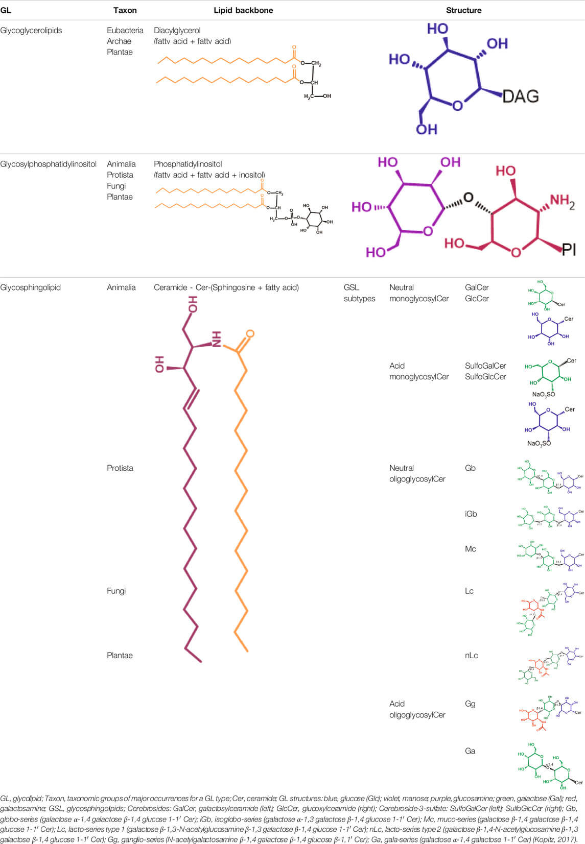 Fig.1 Glycolipid types include glycoglycerolipids, glycosylphosphatidylinositol, and glycosphingolipids.1
The specific sequence of these sugars defines the GSL's identity and function. Globosides are a major family of GSLs in the "globo-series." They are built on a core sugar chain. The two most vital members for researchers are Gb3 and Gb4. The table below summarizes these key targets.
| Target Name(s) | Molecular Identity | Key Binding Partner(s) | Primary Disease Association(s) |
|---|---|---|---|
| Gb3, CD77, Pk Antigen | Globotriaosylceramide | Shiga Toxin (Stx, Verotoxin) | HUS, Fabry Disease, Burkitt's Lymphoma |
| Gb4, P Antigen | Globotetraosylceramide | Human Parvovirus B19 | Fifth Disease, Aplastic Crisis |
Globotriaosylceramide (Gb3): The Toxin Receptor
- Also Known As: CD77, Pk antigen.
- Structure: Ceramide + Glucose + Galactose + Galactose.
- Key Function: Gb3 is most famous as the primary receptor for Shiga toxin (Stx). This is the potent toxin produced by E. coli O157:H7. The toxin binds to the sugar head of Gb3, which allows it to enter and kill the cell.
-
Disease Relevance:
- Hemolytic Uremic Syndrome (HUS): Shiga toxin binding to Gb3 on kidney cells is the direct cause of this life-threatening condition.
- Fabry Disease: This is a genetic lysosomal storage disorder. A faulty enzyme (alpha-galactosidase A) causes Gb3 to build up to toxic levels in cells throughout the body.
- Cancer: Gb3/CD77 is also known to be highly expressed in certain cancers, such as Burkitt's lymphoma, and is involved in signaling cell death (apoptosis).
 Fig.2 Gb3 structure.1
Studying these processes requires tools that can specifically find and bind Gb3. This is why a high-quality anti-Gb3 antibody, anti-CD77 antibody, or anti-PK antigen antibody is so valuable.
Globotetraosylceramide (Gb4): The Viral Gateway
- Also Known As: P antigen.
- Structure: Gb3 + another sugar (N-acetylgalactosamine). Gb4 is essentially an extended version of Gb3.
- Key Function: Gb4 is the primary cellular receptor for Human Parvovirus B19. This common virus binds to the Gb4 sugar chain to infect cells, particularly red blood cell precursors.
- Disease Relevance:
- Parvovirus B19 Infection: Causes "fifth disease" in children, acute arthritis in adults, and severe anemia (aplastic crisis) in immunocompromised patients. The virus's ability to infect cells is entirely dependent on Gb4.
To understand this viral pathway, researchers need a specific anti-Gb4 antibody. This antibody can be used to identify which cells are vulnerable to infection or to develop a parvovirus B19 receptor antibody that can block the virus from binding to these cells.
The Great Research Bottleneck: Why Are Globoside Antibodies So Hard to Make?
If these targets are so important, why are suitable antibodies for them so rare?
The answer lies in their fundamental biology. The immune system is brilliant at recognizing proteins, but it is not well-equipped to "see" lipids. This creates significant technical hurdles for antibody development.
The Lipid-Antigen Problem
Proteins are large, complex, and full of features that trigger a strong immune response. Lipids like Globoside are small. They are considered T-cell independent antigens. This means they often produce a weak, low-affinity immune response, which is typically dominated by IgM antibodies and lacks the robust "memory" and "class-switching" seen in protein responses.
The Membrane-Hiding Problem
Globosides do not float freely. They are embedded in the oily cell membrane, with only their sugar heads exposed. To create a good antibody, you must immunize with an antigen that mimics this natural state. If you just inject purified Gb3, the immune system may make antibodies to the wrong part (like the ceramide tail) or to a shape that doesn't exist on a real cell.
The Purity and Presentation Challenge
Isolating large amounts of pure Gb3 or Gb4 from natural sources is extremely difficult and expensive. The resulting material is a waxy, insoluble lipid. You cannot simply inject this. It must be "presented" correctly, for example, by embedding it into artificial membranes (liposomes) or attaching it to larger carrier proteins. Getting this step right is more art than science, and it is the secret to a successful project.
The Cross-Reactivity Problems
This is the single biggest problem with most commercial anti-glycosphingolipid antibody tools. The cell surface is crowded with dozens of different GSLs that have similar sugar structures.
- Gb4 is built from Gb3.
- Other GSLs (like from the ganglio-series) may share one or two sugars.
An antibody that is not perfectly specific may bind to Gb3, but also to Gb4 and perhaps other molecules. This leads to false positives, confusing data, and unreliable conclusions. A researcher may think they are tracking Gb3, but they are actually tracking three different molecules at once. This is why you need more than just an antibody. You need a custom anti-globoside antibody developed by experts who understand lipid biochemistry and know how to screen for absolute specificity.
Our Solution: A Custom Antibody Service Built for Glycolipids
We are experts in complex antigens. Our custom antibody service is not a generic, one-size-fits-all platform. It is a specialized program designed from the ground up to address the unique challenges of GSLs, such as Gb3 and Gb4. We build the specific tool you need for your exact research question. Our service is a fully collaborative partnership. We work with you at every stage to ensure the final antibody meets your needs.
Step 1: Project Consultation and Antigen Strategy
It all starts with your goal.
- Do you need an anti-Gb3 antibody for Fabry disease research? You will likely need an antibody that works in IHC on tissue samples.
- Do you need a Shiga toxin receptor antibody? You need an antibody that not only binds Gb3 but also blocks the toxin's binding site, likely for use in a cell-based neutralization assay.
- Do you need an anti-CD77 antibody for flow cytometry? You need an antibody that recognizes the native Gb3 on the surface of living cells.
Based on your goal, we design the antigen. We use highly purified or synthetic globosides and conjugate them using proprietary methods to carrier proteins or liposomes. This ensures the immune system sees the globoside in a "cell-like" way, promoting a strong and specific response.
Step 2: Advanced Immunization and Antibody Generation
We offer multiple platforms to generate your antibody, ensuring the best possible outcome.
- Monoclonal Antibody (Hybridoma): This is the classic, gold-standard approach. We immunize animals using our specialized GSL antigen protocols. We then create hybridomas and screen thousands of clones to find the single one that produces the perfect antibody. This results in an eternal, consistent supply of a single, highly characterized antibody.
- Recombinant Antibody (Phage Display): This is a powerful, in vitro alternative. Instead of using an animal's immune system, we use vast libraries of antibody genes (billions of variants). We "pan" this library against the Gb3 or Gb4 target. This allows us to find and isolate human or animal antibody fragments with extremely high affinity and specificity. This method is often faster and allows for more precise engineering.
- Polyclonal Antibody: For some applications, a polyclonal antibody (a mixture of different antibodies binding to different parts of the globoside) is preferred. We can generate high-titer polyclonal serum from immunized animals, which is then purified to isolate only the GSL-specific antibodies.
Step 3: The Most Critical Step: Rigorous, Specificity-Focused Screening
This is what sets our service apart. We do not stop at finding an antibody that binds your target. We identify the antibody that specifically binds to your target. Our screening process is multi-layered:
- Positive Screen (ELISA/Glycan Array): We test all potential antibodies against the purified target.
- Negative/Counter Screen (ELISA/Glycan Array): We then test the "hits" against a panel of closely related GSLs. This panel includes Gb4 (if the target is Gb3), Gb3 (if the target is Gb4), GM1, GD1a, GD1b, ceramide, and other common membrane lipids.
- We discard any clone that shows cross-reactivity.
- Functional Screen (Optional): Based on your project, we can test the final candidates for your application. For example, we can use flow cytometry on cell lines known to express (or not express) CD77. For a parvovirus B19 receptor antibody, we can test its ability to block viral attachment in a cell-based assay.
This rigorous process ensures that the anti-Gb3 antibody you receive binds only to Gb3. It will not bind to Gb4. This gives you data you can trust.
Step 4: Production, Purification, and Final Validation
Once you have selected your final antibody clone, we move to production. We can produce anywhere from milligrams to grams of your custom antibody. It is purified using industry-standard chromatography and passes a final quality control check. You receive a complete validation data package, including its concentration, purity, and the specificity data from our screening cascade. You get a tool that is ready for your experiments, right out of the box.
What You Can Achieve with Our Custom Globoside Antibodies
A truly specific anti-globoside antibody opens doors to research that was previously impossible. Our custom anti-globoside antibody service provides the key to unlocking these complex biological systems.
Cellular Receptor Studies
- Map Receptor Distribution: Use an anti-Gb3 antibody (CD77) or an anti-Gb4 antibody (P antigen) in flow cytometry to quantify precisely what percentage of your cell population expresses the receptor.
- Visualize Receptor Location: Use the antibodies in immunofluorescence (IF) or confocal microscopy to see where on the cell the globosides are. Are they clustered in specific domains (like lipid rafts)?
- Identify Receptor-Expressing Cells: Use immunohistochemistry (IHC) on tissue sections to identify which cell types express Gb3 or Gb4 in complex tissues like the kidney, brain, or bone marrow.
Infectious Disease and Toxinology
- Develop Blocking Antibodies: This is a significant application. We can screen for a Shiga toxin receptor antibody that physically blocks the toxin's binding site on Gb3. This antibody becomes a powerful research tool to study the toxin's mechanism and a potential therapeutic lead.
- Create Viral-Entry Inhibitors: A parvovirus B19 receptor antibody can be used to block the virus from attaching to Gb4 on host cells, effectively neutralizing the virus in cell culture and enabling detailed study of its entry pathway.
- Trace Toxin/Viral Trafficking: By "pre-staining" cells with a fluorescently-labeled anti-CD77 antibody, you can watch in real-time as the Shiga toxin binds and is internalized by the cell.
Fabry Disease Research
- Detect and Quantify Gb3 Buildup: A highly specific anti-Gb3 antibody is the most essential tool for Fabry disease research. It can be used in IHC/IF, ELISA, and Flow Cytometry.
Cancer and Cell Biology
- Identify Cancer Biomarkers: Gb3 (CD77/Pk antigen) is often overexpressed in specific cancers but absent in healthy tissue.
- Study Apoptosis Signaling: Gb3/CD77 is known to be involved in triggering programmed cell death (apoptosis). A custom antibody can be used to probe this pathway, helping to understand how cancer cells may use (or evade) this mechanism.
The Power of Specificity: A Summary of Applications
| Research Field | Antibody Target | Example Experimental Use | Research Question Answered |
|---|---|---|---|
| Toxinology | Gb3 (Shiga Toxin Receptor) | Neutralization Assay | Can we block the toxin from binding? |
| Virology | Gb4 (Parvovirus B19 Receptor) | Flow Cytometry | Which cells are susceptible to infection? |
| Genetic Disorders | Gb3 | IHC on patient tissue | Where is Gb3 accumulating in Fabry disease? |
| Oncology | Gb3 (CD77 / Pk Antigen) | IF on cancer cell lines | Is this cancer targetable via Gb3? |
| Cell Biology | Gb3 / Gb4 | Confocal Microscopy | Where do globosides live on the cell membrane? |
Why Partner with Us?
We are not just a vendor. We are your partner in solving the most difficult challenges in glycobiology.
- We Are GSL Specialists: We focus on the "difficult" targets that generalist companies avoid. We have deep expertise in lipid and carbohydrate chemistry, and we have built our platforms specifically for anti-glycosphingolipid antibody development.
- We Guarantee Specificity: Our reputation is built on our rigorous, multi-step screening process. We deliver antibodies, not "binders." We provide tools tailored to your target, eliminating the risk of cross-reactivity that often plagues this field.
- We Are Fully Custom: We do not sell you an off-the-shelf product. We build a unique antibody tailored to your exact needs, your application, and your research question.
- We Are Transparent: You will be in communication with our scientific team throughout the project. We provide clear milestones, detailed reports, and expert consultation.
Related Services
- Anti-Sulfatide Antibody Development
- Anti-Galactocerebroside Antibody Development
- Anti-Blood Group Antigens Antibody Development
Globoside research is at the forefront of medicine. Understanding these receptors is the key to new therapies for infectious diseases, genetic disorders, and cancer. But you cannot study what you cannot see. Stop letting the lack of specific tools slow down your research. Please tell us your target and application, and we will design a project plan and deliver the high-quality, fully validated custom anti-globoside antibody you need to make the next breakthrough. Contact our team of PhD-level scientists to discuss your project.
Reference:
- Celi, Ana Beatriz, et al. "Role of globotriaosylceramide in physiology and pathology." Frontiers in Molecular Biosciences 9 (2022): 813637. Distributed under Open Access license CC BY 4.0, without modification. https://doi.org/10.3389/fmolb.2022.813637
Supports
- Glycolipid
- GM3 Antibody in Cancer Immunotherapy
- GM3 Ganglioside in Disease & Anti-GM3 Antibody Tools
- Engineering High-Affinity scFv for Next-Generation GD2-CAR-T Therapy
- A Simple Guide to CANOMAD Syndrome
- Anti-Glycolipid Antibody Overview
- GM3 Ganglioside Overview
- Sulfatide and Anti-Sulfatide Antibodies Overview