Custom Anti-TACA Antibody Development Service
Tumor-associated carbohydrate antigens (TACAs) have emerged as crucial targets in cancer research. These glycans are associated with significant alterations in cell behavior, immune recognition, and disease progression. At Creative Biolabs, we support research teams in generating reliable, high-quality anti-TACA antibodies through a focused and efficient development platform, which is also suitable for developing other specialized solutions, such as our Anti-Glycolipid Antibody Development service. Our goal is to provide precise results, steady project management, and reagents you can trust.
What Are the Tumor-Associated Carbohydrate Antigens (TACAs)
Cancer cells often display abnormal surface molecules. These markers fall into three main groups: tumor-associated antigens (TAAs), tumor-specific antigens (TSAs), and tumor-associated carbohydrate antigens (TACAs). TAAs are present in both healthy and tumor tissues, but exhibit higher expression in cancer. TSAs are generally exclusive to tumor cells. TACAs represent a distinct category. They arise from altered glycosylation pathways and are found on the tumor surface in the form of glycoproteins and glycolipids. Their structures are simple, stable, and often repeated in clusters, making them valuable targets for detection and characterization. TACAs are grouped into several major classes. The table below summarizes these categories and links them to our dedicated antibody development services.
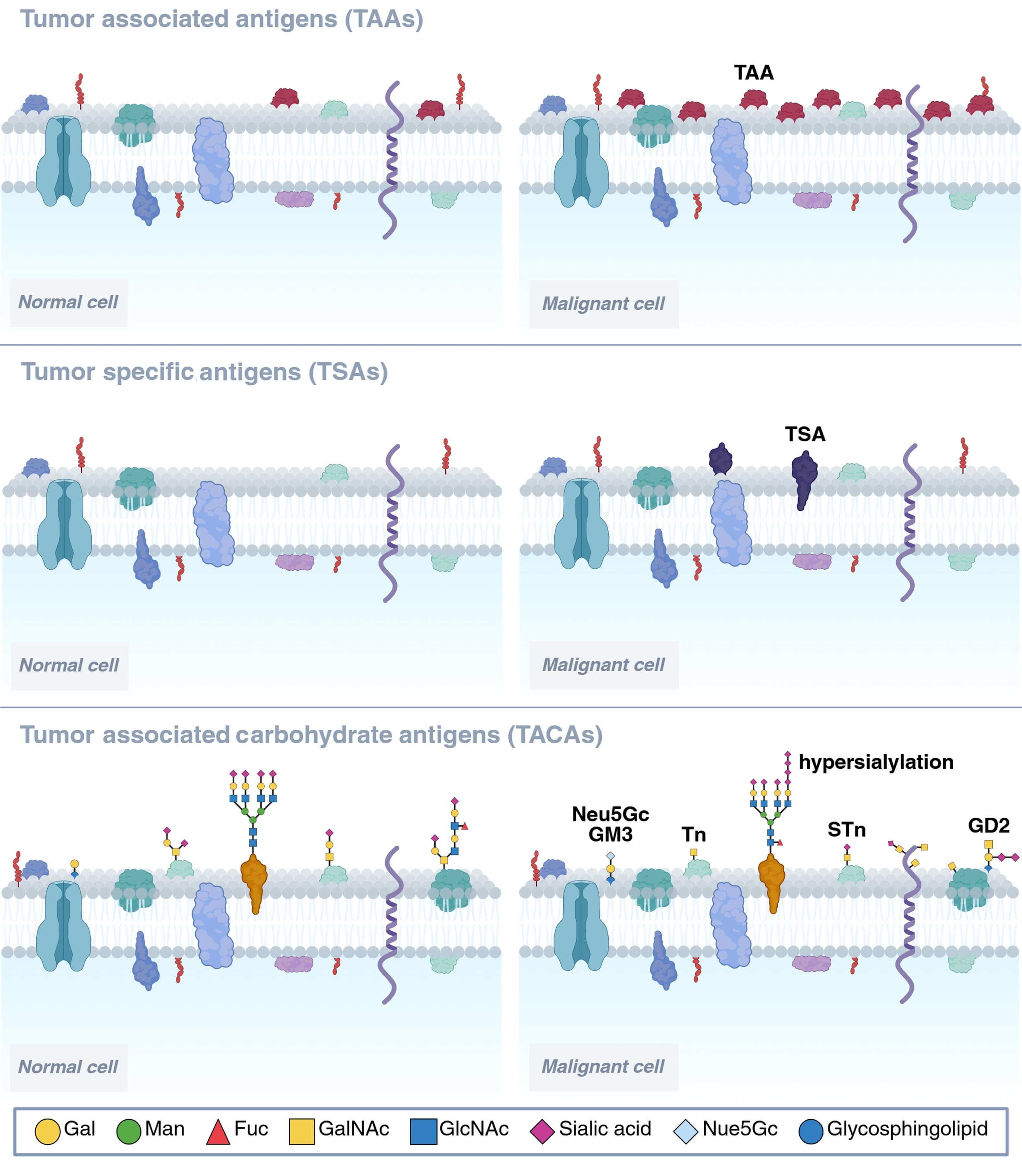 Fig.1 TAAs, TSAs, and TACAs comparison in normal cells and cancer cells.1
Classification of TACAs
| TACA Category | Key Features | Research Focus | Related Service |
|---|---|---|---|
| Tn Antigen | Minimal O-GalNAc structure; frequently exposed in epithelial cancers | Early glycosylation changes and mucin biology | Anti-Tn Antibody Development |
| sTn Antigen | Sialylated Tn, enriched in mucin-producing tumors | Immune modulation and tumor progression | Anti-sTn Antibody Development |
| T / sT Antigens | TF disaccharide and its sialylated form are present in many tumor types | Cell adhesion and metastasis studies | Anti-T/sT Antibody Development |
| sLeA (CA19-9) | Selectin ligand: widely used cancer marker | Biomarker research, selectin-mediated trafficking | Anti-sLeA (CA19-9) Antibody Development |
| sLeX | Selectin ligand tied to cell movement and inflammation | Immune cell and tumor cell migration studies | Anti-sLeX Antibody Development |
| Lewis B | Histo-blood-group-related fucosylated structure | Host–pathogen interaction and cancer profiling | Anti-Lewis B Antibody Development |
| Lewis Y | Overexpressed in several epithelial tumors | Glycan phenotyping and tumor progression studies | Anti-Lewis Y Antibody Development |
Why Anti-TACA Antibodies Matter
Changes in glycosylation are a consistent feature of tumor biology. TACAs influence adhesion, signaling, immune detection, and metastatic spread. They also help researchers distinguish tumors from normal tissue. Reliable anti-TACA antibodies allow clear visualization, accurate quantification, and consistent functional analysis. They support immunostaining, flow cytometry, biomarker assay development, and the use of targeted research tools. Creative Biolabs develops custom anti-TACA antibodies that address the structural precision required to differentiate closely related glycans. Our platform is designed to produce antibodies that function smoothly across multiple applications, providing researchers with reproducible and dependable results.
The Unique Challenge of Developing Anti-TACA Antibodies
Developing anti-TACA antibodies often becomes difficult for research teams. Glycans are weakly immunogenic, and their minor structural differences can lead to cross-reactivity. Many teams also face issues with antigen presentation because TACAs behave differently when displayed on various carriers. These problems often produce antibodies that are unstable, non-specific, or unreliable in downstream assays. At Creative Biolabs, we address these pain points by designing antigens that display the correct glycan density and orientation. This enhances immune recognition and reduces the likelihood of carrier-dominant responses. Our screening strategy includes counter-selection against closely related glycans, helping us isolate clones with strong specificity. Validation takes place using glycan panels, cell surface models, and multiple assay formats, ensuring consistent performance. This approach reduces background signals, enhances affinity, and provides our clients with antibodies that can be used directly in their workflows without further optimization.
End-to-End Workflow for Anti-TACA Antibody Projects
Design Your Anti-TACA Antibody
Custom TACA Antibody Development Services at Creative Biolabs
We can produce IgG, IgM, Fab, scFv, VHH, and recombinant formats tailored to your specific requirements. We also prepare label-ready versions for fluorescent detection, enzyme assays, or biotin systems. Researchers can choose the format that fits IHC, IF, ELISA, flow cytometry, or plate-based screening. Our services include:
Anti-Tn Antibody Development
We design and develop antibodies that recognize the Tn antigen with high accuracy. Tn is a simple O-GalNAc structure often exposed on tumor-associated mucins. Our antibodies enable researchers to study early glycosylation truncation, compare tumor and normal tissue, and support projects in mucin biology and glycan-based classification. We validate performance across IHC, ELISA, IF, and flow cytometry to ensure consistent and reproducible detection.
Anti-sTn Antibody Development
The sTn antigen appears in tumors with heavy mucin secretion and altered sialylation patterns. We generate antibodies that distinguish sTn from related glycans such as Tn or sialylated core structures. These reagents are useful in studies involving tumor invasion, metastasis, and immune cell modulation. Each antibody undergoes counter-screening to confirm that it retains strong selectivity even in complex biological samples.
Anti-T/sT Antibody Development
T and sT antigens serve as markers of glycan exposure that correlate with tumor adhesion and spread. Our antibodies facilitate the investigation of TF antigen biology, observation of glycan-associated adhesion events, and study of changes in glycosylation during tumor progression. These antibodies are suitable for use in cell-based assays, live-cell imaging, and histological evaluation. We offer multiple formats to fit different research workflows.
Anti-sLeA (CA19-9) Antibody Development
sLeA (CA19-9) is commonly used in cancer research as a serum-associated marker and a selectin ligand. We develop antibodies that detect sLeA with high sensitivity and low background. These reagents support quantitative assays, glycan screening, and studies on tumor–endothelial cell interactions. We also provide engineered variants for high-resolution imaging or platform integration.
Anti-sLeX Antibody Development
sLeX is a central molecule in immune cell trafficking and tumor cell movement. Our custom antibodies enable researchers to investigate rolling, adhesion, and cellular positioning in various models. Each antibody is optimized for clear discrimination between sLeX and closely related glycans. We validate function using selectin-based systems and cell assays that measure migration and adhesion.
Anti-Lewis B Antibody Development
Lewis B plays roles in host–pathogen interaction and epithelial glycan regulation. Our team produces antibodies that recognize Lewis B in tissue, cell lines, or purified glycoprotein samples. These reagents help researchers map fucosylation patterns and assess glycan expression in various disease models. We ensure strong specificity by including structured cross-testing with related Lewis family glycans.
Anti-Lewis Y Antibody Development
Lewis Y is re-expressed in many aggressive tumors. We generate antibodies that target this glycan with steady affinity and selectivity. This service supports studies on tumor progression, glycan-based risk assessment, and phenotypic profiling. Researchers often utilize our antibodies in long-term studies due to their stable batch performance and the availability of recombinant production.
Advanced Glyco-Analytical Support
To help clients evaluate glycan-focused antibodies, we provide a wide range of analytical services. This support improves decision-making and accelerates assay development. Our available analytics include:
- Glycoarray platform for glycan specificity profiling
- Glycosylation analysis for protein, antibody, glycolipids, and glycoRNA
- Affinity and kinetics measurement by SPR
Why Researchers Choose Creative Biolabs?
- Strong experience in glycan-focused antibody development
- Detailed antigen design for accurate TACA presentation
- Reliable screening and validation steps
- Full analytic support with glycan-specific tools
- Multiple discovery technologies in one platform
- Consistent data quality and clean documentation
- Ready-to-use antibodies designed for real experimental conditions
How We Work with Your Team?
We support each project with direct communication, predictable timelines, and clear data packages. Our team explains each step and ensures that decisions are made based on the most up-to-date results. You can request adjustments during the project, including additional screening rounds, alternative antigen designs, or extended validation. Our goal is to provide you with a comprehensive and reliable antibody that suits your experiments, eliminating the need for additional troubleshooting. Every project receives dedicated scientific and project management support from start to finish. If you are studying tumor glycosylation, developing new biomarkers, or building a broader cancer research program, Creative Biolabs will help you create precise and dependable anti-TACA antibodies. Share your target, planned applications, and preferred timelines with us. We will deliver a clear project plan and a practical solution that fits your research goals.
Start Your Anti-TACA Antibody Project
Reference:
- Meier, Edward PW, and Andreas H. Laustsen. "Advances in antibody-based strategies for targeting cancer-associated glycopeptide antigens." Drug Discovery Today (2025): 104507. Distributed under Open Access license CC BY 4.0, without modification. https://doi.org/10.1016/j.drudis.2025.104507
Supports
- TACAs Overview
- Guide to Blood Group Antigens
- Comparing sLeA and sLeX Roles in Cancer
- CA19-9 as a Pancreatic Cancer Biomarker
- Lewis Antigen System Overview
- TACA-Targeted ADCs, CAR-Ts, and RICs