Sulfatide and Anti-Sulfatide Antibodies: A Comprehensive Overview
As a leading expert in antibody engineering, Creative Biolabs recognizes that advancements in neuroscience depend on precise tools. Glycolipids, the central targets in many autoimmune neuropathies, are notoriously difficult to study. We are committed to providing a deep introduction to sulfatide and the autoantibodies that target it. To support your research on sulfatide, we offer specialized platforms to build the specific reagents you need, including our Custom Anti-Sulfatide Antibody Service and solutions for other complex glycolipids.
Sulfatide: A Critical Component of the Nervous System
Sulfatide is a type of sulfonated glycosphingolipid. In simpler terms, it is a special fat molecule (a lipid) attached to a sugar (galactose) that has a sulfate group added to it. This molecule is not just an incidental component; it is found in high concentrations in the myelin sheath, which surrounds nerve fibers in both the central and peripheral nervous systems. It plays a vital role in maintaining the structural integrity and function of myelin, as well as supporting neuronal signaling.
What Is the Sulfatide Structure?
The sulfatide structure is what defines its function. It consists of three main parts:
- A Ceramide "Tail": This is a lipid base that anchors the molecule into the cell membrane.
- A Galactose "Head": A sugar molecule that extends from the cell surface.
- A Sulfate Group: This negatively charged group (SO3-) is added to the galactose.
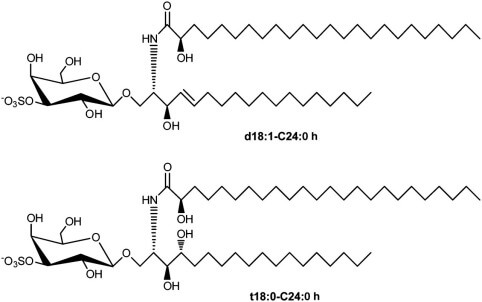 Fig.1 Structure of sulfatide.1
This unique sulfatide structure—with its lipid tail and charged sugar head—allows it to interact with other lipids and proteins, making it essential for myelin's compact and stable organization.
Where is Sulfatide Found?
Sulfatide is incredibly abundant in the nervous system:
- Central Nervous System (CNS): It is a significant component of the myelin sheath produced by oligodendrocytes (which wrap axons in the brain and spinal cord).
- Peripheral Nervous System (PNS): It is also highly expressed in the myelin produced by Schwann cells (which wrap axons in the peripheral nerves).
What is Sulfatide's Function?
Sulfatide is essential for a healthy nervous system. Its primary function is to ensure the stability and function of the myelin sheath.
- Structural Integrity: Sulfatide molecules pack tightly together. Their charged sulfate groups interact with other molecules, helping to hold the multiple layers of the myelin sheath together in a compact, stable structure.
- Maintaining Ion Channels: Sulfatide plays a key role in organizing specific parts of the axon. This is crucial for saltatory conduction—the process that allows nerve signals to jump rapidly down the axon.
- Cell Signaling: It also participates in cell-to-cell signaling and adhesion, helping to guide the interactions between axons and the myelin-producing cells.
When sulfatide is damaged or lost, the entire structure of the myelin sheath can unravel. This leads to "demyelination," where the nerve axon is left exposed. An exposed axon cannot conduct signals properly, leading to the symptoms of autoimmune neuropathy, such as weakness, numbness, and paralysis.
The Research Challenge: Why Studying Sulfatide is Difficult?
At Creative Biolabs, we have spent decades partnering with researchers. We know that studying this process is extremely difficult. Researchers face significant technical bottlenecks that slow down progress, primarily related to the quality of their research tools.
The Antigen is a Lipid
Most antibodies are made against proteins. Standard techniques for antibody development are optimized for protein antigens. Lipids, like sulfatide, are notoriously difficult targets.
- They are small molecules.
- They hide within cell membranes.
- They are difficult to purify and use for immunization.
- They often produce a weak or non-specific immune response.
The Problem of Cross-Reactivity
The nervous system is composed of similar-looking glycolipids (e.g., gangliosides like GM1, GD1a). Many commercially available antibodies are "polyspecific," meaning they accidentally bind to other lipids besides sulfatide. This leads to false positives and confusing data. A study is only as good as its tools, and a non-specific antibody is a poor tool.
Isotype and Subclass Matter
As described in the pathogenic mechanisms, an IgM anti-sulfatide antibody may cause damage differently than an IgG1 or IgG3 antibody. Most research tools do not allow you to study this distinction efficiently. For example, to prove that IgM antibodies cause complement activation, a researcher needs two things:
- A way to detect only IgM-class antibodies in patient samples.
- A purified, pathogenic IgM-class antibody to use in a functional experiment.
These highly specific reagents do not exist off-the-shelf. This is the gap that holds back research.
The Relationship Between Sulfatide and Anti-Sulfatide Antibody
The relationship between sulfatide and anti-sulfatide antibodies is the central event in many forms of autoimmune neuropathy, representing a direct "target-and-attacker" interaction that leads to nerve damage. The target is sulfatide, a typical and essential lipid in the myelin sheath, while the attacker is the anti-sulfatide antibody, an autoantibody from the body's own immune system. In a healthy state, the immune system completely ignores sulfatide. However, in a demyelinating disease state, this tolerance is broken, and the immune system mistakenly identifies sulfatide as a foreign invader, producing and releasing anti-sulfatide antibodies into the bloodstream. These antibodies then circulate until they find their target on the myelin sheath and bind to it tightly. This binding event is the critical trigger for the disease, acting as a "tag" or "flag" that signals the rest of the immune system to "Attack this spot." This initiates a destructive cascade, such as the complement system, which punches holes in the myelin and destroys it. In short, this relationship is the mechanism of the disease, and this specific binding is what translates a simple immune error into a devastating physical attack on the nervous system. Understanding how to block this interaction is, therefore, a key goal for researchers developing new therapies.
Detecting Anti-Sulfatide Antibodies (For Research Use Only)
A critical part of studying these diseases is the detection of anti-sulfatide antibodies in research samples. This is often accomplished using immunoassays. In a laboratory setting, a sulfatide antibody test is typically an ELISA (Enzyme-Linked Immunosorbent Assay). The basic principle involves several key steps:
- Antigen Coating: A purified sulfatide preparation is used to coat the bottom of a microplate well.
- Sample Incubation: A research sample (e.g., serum from an animal model) is added to the well.
- Binding: If anti-sulfatide antibodies are present in the sample, they will bind to the sulfatide coated on the plate.
- Detection: A secondary antibody, which binds to the first antibody and carries an enzyme, is added. This enzyme produces a color change, which can be measured.
This same principle applies to what might be called a sulfatide autoantibody test or an anti-sulfatide antibody blood test in the context of pre-clinical research—they are all methods to quantify the presence of these antibodies in a given sample to understand disease models better. The accuracy of this test depends on the purity of the sulfatide antigen and the specificity of the detection antibodies. This is why the research challenges mentioned earlier (cross-reactivity, lipid antigens) are so critical to overcome.
Discover Your Specially Designed Anti-Sulfatide Antibodies
Build Your Breakthrough Tool: Our Custom Antibody Capabilities
Your research is unique. Your tools should be, too. We don't just offer a catalog; we provide a fully customized development platform to build the precise anti-sulfatide antibody your research demands. You define the project. Need a high-affinity rabbit monoclonal for unmatched specificity? Or a specific pathogenic isotype, like IgM, to model disease mechanisms in vitro? We can deliver. Our expertise extends to llama VHH and other species to fit your exact experimental design. We overcome the challenges of lipid targets by using advanced antigen strategies, including sulfatide liposomes and carrier conjugates, to ensure a robust and specific immune response. Finally, get your antibody ready-to-use, saving you months of optimization. We deliver in any format, from full-length antibodies to smaller Fab fragments. We can also provide direct labeling with Biotin, HRP, or FITC. We then validate your custom antibody in your specific application—whether ELISA, IHC, or flow cytometry—and guarantee its performance.
Applications: How Your Custom Antibody Accelerates Research
By ordering a custom-built antibody, you move from "making do" with existing tools to designing the perfect tool for your question.
Application 1: Developing Advanced Research Assays
"How can I reliably detect and quantify different isotypes of anti-sulfatide antibodies in biological samples from my disease models?"
Our Solution: We can develop a highly specific rabbit anti-sulfatide antibody that has been validated for ELISA. You can use this antibody as the "capture" reagent to build a robust new research assay.
- Coat an ELISA plate with our custom rabbit antibody.
- This antibody "captures" all the sulfatide from a purified solution.
- Add your research sample (e.g., serum from an animal model or samples from a research biobank).
- If the sample contains a sulfatide antibody, it will bind to the captured sulfatide.
- You can then detect the isotype (e.g., IgM or IgG) using a standard secondary antibody.
This custom-built research tool will be far more specific and sensitive than assays built on non-specific lipid antigens, providing you with more reliable and accurate data for your pre-clinical studies.
Application 2: Unraveling Pathogenic Mechanisms (In Vitro)
"Does the IgM-class anti-sulfatide antibody directly cause complement-mediated damage to oligodendrocytes?"
Our Solution: Order a custom mouse monoclonal IgM anti-sulfatide antibody.
- Grow oligodendrocytes (myelin-producing cells) in a dish.
- Add your custom IgM antibody to the culture.
- Add a source of complement (e.g., normal human serum).
- Observe the results. You can stain for complement deposition (C3b, C5b-9) and cell death.
- Control: Use a non-binding IgM monoclonal to prove the effect is specific to sulfatide binding.
This experiment, which is impossible without a custom reagent, directly answers a fundamental question about disease pathogenesis.
Application 3: Visualizing Myelin Damage in Tissue
"In my animal model of demyelinating disease, is the loss of sulfatide happening before or after the nerve is damaged?"
Our Solution: We will develop a custom rabbit anti-sulfatide antibody that is guaranteed to work in Immunohistochemistry (IHC) on fixed nerve tissue.
- Take tissue sections (brain, spinal cord, or peripheral nerve) from your disease model animals at different time points.
- Stain one section with your custom anti-sulfatide antibody (to see the myelin).
- Stain an adjacent section with an axon marker (like neurofilament).
- By comparing the images, you can precisely map where and when sulfatide is lost relative to the axon itself.
Sulfatide is a vital component of the nervous system, and anti-sulfatide antibodies are key players in the destructive autoimmune response of many demyelinating diseases. Ideas no longer limit progress in understanding these conditions, but by the availability of precise, reliable, and specific research tools. Standard reagents often fail to meet the challenges posed by lipid antigens and the need for absolute specificity. As your dedicated partner in research, Creative Biolabs is committed to providing the custom tools you need to get clear, publishable, and transformative results. Contact us for more service details!
Reference:
- Takahashi, Tadanobu, and Takashi Suzuki. "Role of sulfatide in normal and pathological cells and tissues." Journal of lipid research vol. 53,8 (2012): 1437-50. Distributed under Open Access license CC BY 4.0, without modification. https://doi.org/10.1194/jlr.R026682