Lewis Antigen System: A Comprehensive Overview
While most people are familiar with the ABO and Rh blood groups, our cells are coated in many other compLeX glycans, with the Lewis antigens being among the most critical. The Lewis system is a family of histo-blood group antigens. Unlike ABO antigens, Lewis antigens are primarily found on epithelial tissues and in bodily secretions. Their presence on red blood cells is secondary; they are produced elsewhere and adsorb onto the cell surface from plasma. This family includes Lewis A (LeA), Lewis B (LeB), Lewis X (LeX), Lewis Y (LeY), and the well-known cancer-associated carbohydrate antigens Sialyl-Lewis A (sLeA or CA19-9) and Sialyl-Lewis X (sLeX). These molecules play critical roles in various biological processes, including susceptibility to infectious diseases and cancer metastasis. However, studying the Lewis system poses a significant research challenge due to the antigens' structural similarity, compLeX genetics, and difficulties in detection. This guide from Creative Biolabs explores the Lewis antigen family, including how they are formed, their biological functions, and why they are critical targets for modern research.
How Lewis Antigens Are Made?
You cannot understand the Lewis antigens without first understanding the two key genes that build them: FUT2 and FUT3. These genes do not code for the antigens themselves. They code for enzymes called fucosyltransferases (FUTs). These enzymes are like molecular construction workers. Their only job is to grab a specific sugar molecule, fucose, and attach it to a particular spot on a growing glycan chain. The final structure built by these enzymes depends on two things:
- The precursor chain (the foundation)
- Which enzymes are active (the workers on site)
The Precursor Chains: Type 1 vs. Type 2
The "foundation" for all Lewis antigens is a simple two-sugar chain. But this chain comes in two slightly different forms:
- Type 1 Chain: Galactose linked to GlcNAc in a β1-3 linkage. (Found in secretions, on epithelial cells, and in plasma)
- Type 2 Chain: Galactose linked to GlcNAc in a β1-4 linkage. (Found on all cells, including red blood cells)
This simple difference in linkage is the critical branch point that separates the Lewis family.
- LeA and LeB are built on Type 1 chains.
- LeX and LeY are built on Type 2 chains.
The Enzymes: FUT3 (Lewis) and FUT2 (Secretor)
- FUT3 (The Lewis Gene): This gene produces the "Lewis enzyme." This enzyme adds a fucose molecule to the GlcNAc sugar. If this gene is active, a person is "Lewis-positive." If it is inactive (mutated), they are "Lewis-negative."
- FUT2 (The Secretor Gene): This gene produces the "Secretor enzyme." This enzyme adds a fucose molecule to the Galactose sugar. If this gene is active, a person is a "Secretor," meaning they secrete ABH antigens into their saliva and bodily fluids. If it is inactive, they are a "Non-secretor."
The interplay between these two genes gives rise to the primary Lewis phenotypes.
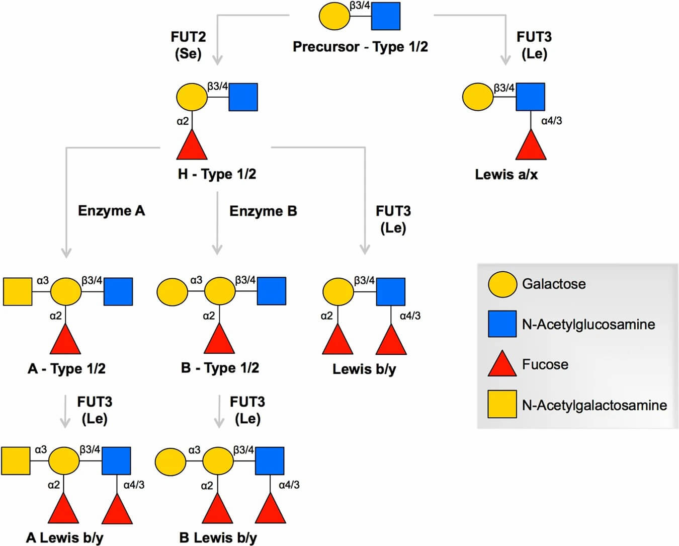 Fig.1 Biosynthetic pathway of ABH and Lewis blood group antigens.1
The Result: The Four Lewis Phenotypes
| Phenotype | FUT3 (Lewis) Status | FUT2 (Secretor) Status | Result / Description |
|---|---|---|---|
| Le(a+b-) / Non-Secretor | Active | Inactive | The active FUT3 enzyme adds fucose to the Type 1 chain, creating the Lewis A (LeA) antigen. Because the FUT2 enzyme is inactive, no H-antigen is made on this chain, and LeB cannot be formed. These individuals are Non-secretors and have LeA antigen in their plasma and on their cells. |
| Le(a-b+) / Secretor | Active | Active | The active FUT2 enzyme first adds fucose to the Type 1 chain, creating the H-antigen. The active FUT3 enzyme then adds a second fucose to that same chain, completing the Lewis B (LeB) antigen. The FUT2 enzyme is more efficient, so almost all Type 1 chains are converted to LeB, Leaving very little LeA behind. This is the most common phenotype. |
| Le(a-b-) / Lewis-Negative | Inactive | Can be active or inactive | Because the FUT3 (Lewis) gene is inactive, the enzyme needed to create both LeA and LeB is missing. These individuals cannot make either antigen, regardless of their Secretor status. |
| Le(a+b+) | (Varies) | (Varies) | This phenotype is very rare in adults (in Caucasians) but is common in infants as the system matures. It is also more common in some Asian populations. |
The Lewis Antigens Introduction
- The simple Lewis antigen, built on a Type 1 chain with a single fucose added by FUT3.
- Primarily known as the antigen of "non-secretors." It can serve as an attachment point for some pathogens. Its presence (or absence) can define susceptibility to certain infections.
- The compLeX Type 1 antigen, with two fucose molecules added by both FUT2 and FUT3.
-
As described, LeB is not made by red blood cells. It is produced by epithelial cells, secreted into plasma, and passively adsorbs onto the red blood cell surface. Its primary expression is in:
- Epithelial Tissues: The lining of the stomach, intestines, and respiratory tract.
- Exocrine Glands: Salivary glands.
- Bodily Secretions: Saliva, milk, and gastric juice (in Secretor individuals).
-
LeB plays a crucial role in the host-pathogen interaction:
- Helicobacter pylori: This is the most famous example. H. pylori is a bacterium that causes chronic gastritis, stomach ulcers, and is the strongest known risk factor for gastric cancer. Its surface protein, BabA, binds explicitly to LeB on stomach epithelial cells. This binding is the critical first step that allows the bacteria to colonize the stomach.
- Norovirus: The LeAding cause of viral gastroenteritis. Many strains of norovirus recognize and bind to LeB (and other blood group antigens) on gut epithelial cells to initiate infection. An individual's blood group antigen profile can determine their susceptibility to different norovirus strains.
- Candida albicans: This opportunistic fungus also shows binding interactions with LeB.
- In healthy adults, LeB expression is restricted. However, in many types of cancer, it is re-expressed at very high levels, making it a Tumor-Associated Carbohydrate Antigen (TACA). High levels are found in colorectal, pancreatic, gastric, and ovarian cancers, and their presence is often linked to increased cell motility.
- To study H. pylori binding, a researcher must have an antibody that only sees LeB and not the highly similar LeA or LeY antigens. A cross-reactive antibody would give false-positive results. This requires tools of extreme precision, often demanding a Custom Anti-Lewis B (LeB) Antibody Development service to create and validate an antibody for this specific functional purpose.
- The Type 2 chain analog of LeA. It's a Type 2 chain with a single fucose added by enzymes like FUT4, FUT7, or FUT9.
- LeX is most famous under another name: SSEA-1 (Stage-Specific Embryonic Antigen 1). It is a classic marker for pluripotent stem cells, including mouse embryonic stem cells and induced pluripotent stem cells (iPSCs). As these cells differentiate, they lose LeX expression. This makes anti-LeX antibodies a fundamental tool in any stem cell research lab.
- Like its Sialylated cousin, LeX is involved in cell-cell recognition and adhesion, particularly in the brain and immune system.
- The Type 2 chain analog of LeB. It's a Type 2 chain that has been "fucosylated" twice.
- While LeB is a TACA, LeY is arguably an even more significant one. LeY is massively overexpressed in a considerable number of human cancers, including 70-90% of breast, lung, ovarian, colorectal, and pancreatic carcinomas.
- In healthy tissue, LeY is restricted. In cancer, its expression "explodes." This is not just a side effect; LeY is an active participant. It integrates into signaling pathways and is believed to promote tumor cell proliferation, survival, and resistance to chemotherapy.
- This makes LeY one of the most attractive TACA targets for new cancer therapies, including antibody-drug conjugates (ADCs) and CAR-T cells. Developing these therapies is entirely dependent on having a particular antibody, which is why Custom Anti-Lewis Y (LeY) Antibody Development is a significant focus for oncology research.
- The LeA antigen with a sialic acid cap.
- As the pancreatic cancer biomarker.
- Sialyl-Lewis A is known worldwide by another name: CA19-9 (Carbohydrate Antigen 19-9). CA19-9 is the gold-standard tumor marker for pancreatic cancer. In a healthy person, levels of CA19-9 in the blood are very low. In a patient with pancreatic adenocarcinoma, these levels can be astronomically high. It is the primary tool used by oncologists to monitor a patient's response to treatment and to screen for disease recurrence.
- It is also elevated in other gastrointestinal cancers, like gastric and colorectal cancer.
- The critical need in this field is for more sensitive and more specific detection assays. Developing the next generation of diagnostic tools relies entirely on sourcing better, higher-affinity antibodies, a challenge addressed by Custom Anti-sLeA (CA19-9) Antibody Development programs.
- The LeX antigen with a sialic acid cap.
- sLeX is the primary functional ligand for a family of proteins called Selectins (E-selectin, P-selectin, L-selectin).
- This "selectin-sLeX" interaction is one of the most-studied mechanisms in cancer biology. Blocking it is a "holy grail" for preventing metastasis.
- Researchers in this field need antibodies that can specifically recognize sLeX to study this process, or functional-grade antibodies to try to block it. This is the goal of Custom Anti-sLeX Antibody Development.
Lewis Antigens in Serological and Immunological Research
The Lewis system is a foundational model in glycobiology for studying human antibody responses to carbohydrate antigens. The focus for researchers is often on the formation and unique biochemical properties of Lewis antibodies, which are distinct from many protein-based antibody responses. For further research, labs require a constant supply of high-quality, reliable typing reagents. This work requires antibodies that can definitively identify Lewis antigens on red blood cells, a core challenge in creating tools for transfusion diagnostics. This need for reliable reagents is a key part of the Custom Anti-Blood Group Antigens Antibody Service, which aims to produce these essential tools for both common and rare blood types.
A Research Model for Antibody Formation
Individuals with the Le(a-b-) phenotype are of significant interest to immunologists and serologists. Because their bodies do not produce LeA or LeB, they provide a valuable human research model for understanding how the immune system generates antibodies (such as anti-LeA or anti-LeB) in response to foreign glycan exposure.
Investigating Antibody Binding Properties
From a biochemical perspective, Lewis antibodies are a classic subject for studying temperature-dependent antibody-antigen interactions.
- The majority of Lewis antibodies are of the IgM isotype.
- A key characteristic studied by researchers is their cold-reactive binding. These antibodies show strong agglutination or binding activity at room temperature or below.
- Crucially, this binding activity is often lost or significantly reduced at 37°C. This property makes them essential subjects for studying the thermodynamics of antibody-glycan binding.
A Complicating Factor in Serological Research
For researchers analyzing serum or plasma samples, these high-titer, cold-reactive IgM antibodies can be a significant technical complication. In an assay, they can cause strong background binding that may obscure the detection of other, lower-affinity antibodies or different research targets. This interference makes it difficult to validate serological assay results, a common challenge in immunological research.
Advance Your Lewis Antigen Research
The Lewis antigens are far more than a minor blood type. They are a perfect example of how compLeX carbohydrates run our biology.
- They form the docking sites for major human pathogens (LeB).
- They are the master switches of embryonic development (LeX).
- They are the billboards that signal cancer (LeY, sLeA).
- They are the functional tools that cancer uses to spread (sLeX).
From the blood bank to the cancer clinic, these molecules are at the center of critical research. However, as this guide has shown, the structural compLeXity, high similarity, and low immunogenicity of these glycans make them incredibly difficult to study. Sourcing an antibody with proven, monospecific binding is the single most significant bottleneck for researchers. This is the exact challenge Creative Biolabs is built to solve. We specialize in overcoming the unique hurdles of anti-glycan antibody development. Where others fail due to cross-reactivity, our advanced platforms succeed. We design immunization strategies to break tolerance and validation cascades, ensuring specificity. Your research is too important to be stalled by a bad antibody.
- If you are studying cancer metastasis, explore our Custom Anti-sLeX Antibody Development and Custom Anti-sLeA (CA19-9) Antibody Development services.
- If you are targeting tumor signaling or host-pathogen binding, see our Custom Anti-Lewis Y (LeY) Antibody Development and Custom Anti-Lewis B (LeB) Antibody Development programs.
Contact our scientific team today. Let's discuss your project and build the precise, validated antibody you need to drive your discovery forward.
Reference:
- Barbé, Laure, et al. "Histo-blood group antigen-binding specificities of human rotaviruses are associated with gastroenteritis but not with in vitro infection." Scientific reports 8.1 (2018): 12961. https://doi.org/10.1038/s41598-018-31005-4
Related Services
Anti-Glycolipid Antibody Development
Anti–Blood Group Antigens Antibody Development
Anti-sLeA (CA19-9) Antibody Development
Anti-sLeX Antibody Development
Anti-Lewis B (LeB) Antibody Development
Anti-Lewis Y (LeY) Antibody Development
Supports
- Glycolipid
- Blood Group Antigen
- Understanding Glycosylation
- Glycosylation Influences Blood Type
- TACAs Overview
- Guide to Blood Group Antigens
- Comparing sLeA and sLeX Roles in Cancer
- CA19-9 as a Pancreatic Cancer Biomarker
- Lewis Antigen System Overview
- TACA-Targeted ADCs, CAR-Ts, and RICs