Custom Anti-GD2 Antibody Service for Neuroblastoma & Cancer Research
Ganglioside GD2 serves as a primary drug target and preclinical imaging probe which spans neuroectodermal tumor cell membranes while providing an established high-value tool for mechanism studies, target validation, imaging, and combination screening applications in oncology research. Enriched on the surface of neuroblastoma, as well as melanoma and sarcoma, GD2 is a tumor-associated carbohydrate antigen that presents abundant, membrane-embedded epitopes for binding studies and functional assays. But carbohydrate epitope recognition is defined by a precise three-dimensional fit—often dictated by "end-on" saccharide insertion in a solvent-structured pocket—so specificity must be engineered and proven, not assumed. At Creative Biolabs, we design and manufacture anti-ganglioside antibody tools from discovery and validation to scalable production, which give researchers rigorous, reproducible ways to interrogate the GD2 antigen in cell and tissue systems. Our programs include mouse, rabbit, and camelid repertoires; hybridoma, phage display, and single B-cell routes; and assays suite tuned to the unique properties of glycolipid targets. For a faster start to your project, we offer a range of off-the-shelf anti-GD2 antibody products.
The GD2 Antigen: A Prime Target in Oncology
To appreciate the power of an anti-GD2 antibody, we must first understand the target itself. GD2 is a glycolipid (a lipid with a carbohydrate attached), and more precisely a disialoganglioside (glycolipid with two residues of sialic acid) that resides in the cell membrane. GD2 is present in human cells and contributes to some essential, but mostly minor, functions especially during fetal development of the nervous system. Expression of GD2 antigen in adult tissues is strictly limited, but exceptions are different tumors of neuroectodermal origin, where the antigen is massively upregulated. As such, tumors where GD2 is highly expressed on the surface include:
- Neuroblastoma (present in >95% of cases)
- Melanoma
- Small-Cell Lung Cancer (SCLC)
- Sarcomas (Ewing sarcoma, osteosarcoma)
- Glioblastomas
The high levels of tumor-associated expression and the limited expression on normal tissues (predominantly on peripheral nerve fibers and cerebellar Purkinje cells) provides a high therapeutic index, since cancer cells can be targeted with limited off-target toxicities to the rest of the body (a mainstay in the field of oncology). Moreover, GD2 has been found to be an active participant in the promotion of cancer, since its binding to cell surface proteins can affect many of the well-known hallmarks of cancer such as cell adhesion, migration and proliferation.
What You Can Do with a GD2 Antibody in Research
- Map GD2 distribution in panels of tumor cell lines and xenograft tissues, quantify receptor-proximal effects, and track membrane biology with high contrast.
- Build different ganglioside GD2 antibody formats for neuroblastoma therapy mechanism studies strictly in preclinical systems, for instance, you can bulid custom high-affinity GD2 scFv for GD2-CAR-T therapy.
- De-risk cross-reactivity versus GD3/GM2 and other TACAs using orthogonal assays and glycan panels to ensure signal fidelity in complex samples.
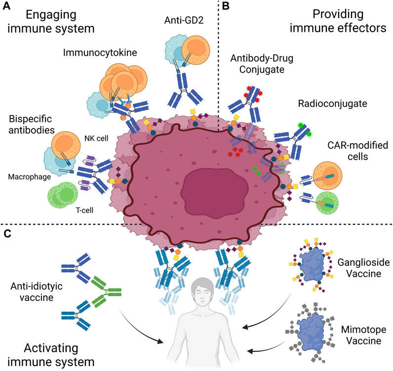 Fig.1 GD2-targeted immunotherapy strategies overview.1
Anti-GD2 Antibody Development Service at Creative Biolabs
We create and rigorously confirm anti-GD2 reagents with native behavior and desired robustness: we present the antigen of interest in two ways (GD2-carrier conjugates and GD2-liposomes) to select for the native epitope, perform hybridoma, phage display, and single B-cell technologies to ensure we find the elusive paratope, use GD3/GM2 counterselective screens from discovery to ensure we are policing specificity, and triangulate data with ELISA , BLI/SPR, flow, IHC/IF, and RUO effector assays; lastly we customize the format you need (IgG, Fab, scFv, VHH with optional labels or Fc edits), manufacture to scale by affinity purification with full QC (purity/aggregate/endotoxin), and provide a ready validation package so your GD2 antibody can move forward confidently in your neuroblastoma-related research programs that target the GD2 antigen.
Our End-to-End Workflow
- Project scoping → target definition, desired format (IgG, Fab, scFv, VHH), assay priorities, and counter-antigen list.
- Antigen build → GD2 conjugates and liposome presentation engineered and qualified.
- Immunization & library creation → species strategy (mouse/rabbit/camelid) aligned to epitope goals.
- Primary screening → parallel ELISAs (conjugate/liposome) and competitive binding.
- Specificity gate → glycan array & cross-panel negative selection (GD3, GM2, GM3, Globo).
- Kinetic and cell-based validation → BLI/SPR, flow cytometry, IHC/IF.
- Engineering & polishing → reformatting, Fc edits, conjugations.
- Scale-up & QC → affinity purification, release testing, and documentation package.
Typical Applications in Research
We deploy GD2 antibody tools to answer four practical questions—how much GD2 antigen is on your cells or tissues, what a candidate anti-GD2 antibody can do functionally in vitro, how clean its specificity is across related gangliosides, and whether the reagent remains stable and comparable across formats and lots.
| Your Research Question | Our Approach | What You Receive |
|---|---|---|
| "How strongly does my antibody bind to its target?" | We use advanced methods like Flow Cytometry and BLI/SPR. | A clear report with binding affinity data and antigen levels. |
| "Does my antibody actually kill cancer cells?" | We perform key functional studies like cell-killing assays (ADCC/CDC). | Easy-to-read graphs showing your antibody's cell-killing performance. |
| "How specific is my antibody to GD2?" | We test it against a panel of similar molecules using ELISA and Glycan Arrays. | A definitive answer on your antibody's specificity and cross-reactivity risks. |
| "Where is the GD2 target in my tissue samples?" | We use standard tissue staining techniques (IHC/IF). | High-quality images showing the precise location of GD2 in your tissues. |
| "Can you label my antibody for detection assays?" | We conjugate your antibody with fluorescent dyes, biotin, or other labels. | A ready-to-use labeled antibody, quality-controlled for your specific assay. |
| "Can I get the sequence for engineering a CAR-T?" | We sequence the antibody and map its binding site (epitope binning). | The complete antibody sequence, ready for your cell engineering program. |
| "How does my antibody work with other drugs?" | We design and run co-treatment studies with other agents. | A synergy report showing if the drug combination is more effective. |
| "Is the antibody stable and reproducible?" | We conduct stability studies and rigorous batch-to-batch comparisons. | A full report proving your antibody's stability and lot-to-lot consistency. |
Ready-to-Use Antibody Targeting GD2
- Prefer an off-the-shelf start? Visit the ready-to-use GD2 antibodies & reagents options page above to select a product that fits your use case.
- If your study requires a different host, format, or label, or extended validation, please contact us for a custom anti-GD2 antibody build.
- Need a side-by-side? We routinely run catalog vs. custom comparability tests under identical conditions (RUO) and share the raw plots.
| Criterion | Catalog (Ready-to-Use) | Custom Program |
|---|---|---|
| Timeline | Immediate availability | Built to spec (project-based) |
| Format needs | Standard formats/labels | Any format (IgG/Fab/scFv/VHH, Fc edits) |
| Specificity profile | Fixed, documented | Tunable; add GD3/GM2 counterscreens |
| Assay depth | Basic validation | Full validation matrix & dossier |
| Scale | Pre-set sizes | Fit-for-purpose, from mg to multi-grams |
The Challenge with Glycolipid Targets—and How We Solve It
Problem 1: It's hard to get a good immune response against GD2.
GD2 don't trigger a strong immune reaction by themselves. To solve this, we attach GD2 to a larger carrier protein or place it in a structure that mimics a cell membrane. This approach creates a much stronger response. We make both versions and test them side-by-side to find the best one for your project.
Problem 2: GD2 looks very similar to other related molecules.
Because other molecules like GD3 and GM2 are structurally similar to GD2, an antibody might bind to the wrong target. Our solution is to screen for specificity right from the start. We run tests to actively weed out any antibodies that bind to these related molecules, ensuring high precision.
Problem 3: The antibody's binding needs to be perfect.
Recognizing sugar molecules is tricky. The fit between the antibody and GD2 must be exact, and even tiny structural changes can cause the antibody to fail. To solve this, we use existing 3D structural data of GD2-antibody complexes. This information guides our design process, helping us select antibodies with the perfect structure for precise binding.
Popular Services You May Be Interested in
- Anti-GD1a Antibody Development
- Anti-GD1b Antibody Development
- Anti-GD3 Antibody Development
- Anti-Ganglioside Antibody Development
- Glycosphingolipids Analysis Service
- Glycan Microarray Profiling
Share your brief: intended application, desired format, control antigens, assay preferences, and scale. We will propose an antigen plan, discovery route, validation matrix, and a documentation package aligned to your downstream needs. We are committed to providing you with a concrete, membrane-aware plan for a GD2 antibody or anti-GD2 antibody set—optimized for your models, and documented for reproducibility. Contact our experts today to get your custom anti-glycolipid antibody design service!
Reference:
- Machy, Pierre, Erwan Mortier, and Stéphane Birklé. "Biology of GD2 ganglioside: implications for cancer immunotherapy." Frontiers in Pharmacology 14 (2023): 1249929. Distributed under Open Access license CC BY 4.0, without modification. https://doi.org/10.3389/fphar.2023.1249929