Antigen-associated Glycolipid Analysis Service
Introduction
A glycolipid molecule has two parts: a lipid tail and a carbohydrate head. But what does it take to be an antigen? It's all about that glycan chain, which is the epitope—the molecular shape that an immune cell such as a B cell or T cell and its antibody product can recognize. The immune system can initiate a strong response when it identifies glycolipid epitopes as foreign (present on bacteria) or abnormal (found on cancer cells). This is an example of antigen glycosylation—when the carbohydrate component of a molecule gives antigenicity. Creative Biolabs has a suite of glycolipid analysis solutions and a team of highly experienced specialists, who are here to provide you with the high-quality data you need to progress your research, enabling your research to develop better diagnostic and therapeutic strategies.
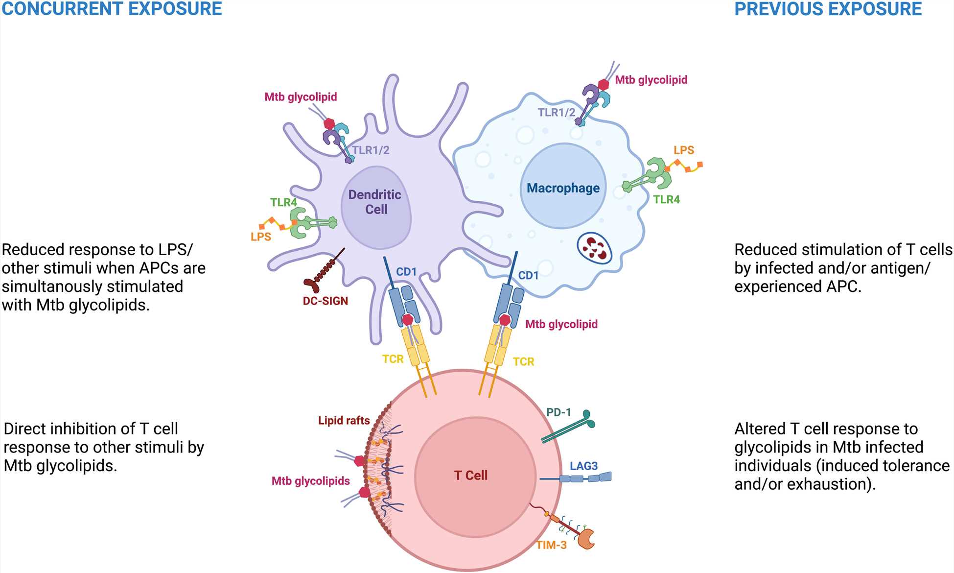 Fig.1 Mycobacterial glycolipids and immune hyporesponse.1
Common Glycolipid Antigens
Glycolipid Blood Group Antigens
One of the most classic examples of glycolipid antigens is the ABO blood group system. The A, B, and H antigens that determine your blood type are not proteins, but carbohydrate structures present on glycolipids (and glycoproteins) on the surface of red blood cells. Your immune system produces antibodies against the blood group antigens you don't have. This is why blood type matching is critical for transfusions—a mismatch leads to a massive immune attack on the transfused cells, triggered by these glycolipid antigens.
Mycobacterial Glycolipid Antigens
The cell wall of Mycobacterium tuberculosis (the bacterium that causes tuberculosis) is rich in unique glycolipids. These molecules, such as Lipoarabinomannan (LAM) and Trehalose Dimycolate (TDM), are essential for the bacterium's structural integrity and its ability to modulate the host immune response. However, they also serve as powerful antigens, stimulating both antibody and T-cell responses.
Know About Our Microarray for Microbial Glycan Antigen
Phenolic Glycolipid Antigens (PGLs)
Another class of mycobacterial lipids, phenolic glycolipid antigens, like PGL-1 from Mycobacterium leprae (the cause of leprosy), are highly specific and immunogenic. The unique sugar composition of PGL-1 makes it a prime target for diagnostic tests, as antibodies against it are found almost exclusively in patients with leprosy.
Our Approach to Antigen-Associated Glycolipid Analysis
At Creative Biolabs, we see the analysis of a glycolipid antigen as telling a complete scientific story. It starts with identifying a molecule of interest on a cell surface and ends with definitively proving it triggers an immune response. Our services are designed to connect every chapter of that story into a single, seamless narrative, including:
- Glycolipid antigen analysis as a complete scientific narrative
- Identifying targets via differential glycolipid profiling (healthy/diseased, pathogenic/harmless)
- Highlighting candidates using patient antibody probes
- Purification ensuring activity originates solely from the glycolipid
- Defining precise identity: molecular formula, glycan sequence, and 3D epitope (MS/NMR)
- Confirming antigenicity: patient antibody binding detection (custom glycolipid microarray)
- Quantifying antigen potency via interaction kinetics (SPR)
Accepted Samples
The quality of every downstream analysis depends on the purity of the starting material. Our team possesses deep expertise in extracting and profiling glycolipids from a vast array of challenging biological matrices. We handle different source materials, including but not limited to:
- Mammalian tissues (Brain, Spleen, etc.)
- Cultured cell lines (Adherent & Suspension)
- Biofluids (Serum, Plasma, Cerebrospinal fluid)
- Bacterial & Fungal pellets
- Plant tissues
Technologies We Used for Glycolipid Analysis
Glycolipid Structural Characterization
This is the core of our analytical power. We use an orthogonal approach, combining multiple cutting-edge technologies to leave no ambiguity in the antigenic glycolipid structure. Our services include MS and NMR. MS precisely determines a glycolipid's molecular formula, sequences its structure, and visualizes its exact location in tissues. While MS provides the "what," NMR provides the "how it's connected." For novel structures or when absolute certainty is required, NMR is the undisputed gold standard that provides a unique spectral fingerprint or 3D structure for a purified glycolipid.
Antibody & Autoantibody Interaction Analysis
A structure is only a blueprint; its function as an antigen must be experimentally validated. Our services are designed to measure and quantify the very interactions that define a glycolipid as an antigen.
- Custom Glycolipid Arrays: The premier tool for screening. We can immobilize your purified glycolipid or use our pre-built library of >100 biologically relevant glycolipids (including a full panel of gangliosides, sulfatides, and bacterial antigens) onto a microarray chip. You can then screen patient/animal serum or purified antibodies to quantitatively map binding profiles, identifying crucial glycolipid antigens and autoantibodies.
- ELISA: The classic workhorse for validating a specific antigen-antibody interaction.
- SPR / BLI: These label-free, real-time technologies provide precise kinetic data, allowing you to measure the affinity and stability of an antibody-glycolipid interaction.
We're here to support your research and help you explore the potential of glycolipid antigen analysis. For more details on our antigen-associated glycolipid analysis service, please feel free to contact our team.
FAQs
Q: What problems does your antigen-associated glycolipid analysis service actually solve for my program?
A: We connect structure to immune function. Our workflow profiles complex glycolipids, defines antigenic epitopes, and quantifies antibody/T-cell interactions. You receive unambiguous structures, ranked antigen candidates, and validated binding/kinetic data—so you can decide which glycolipid antigens merit diagnostic assay development, immunogenicity risk assessment, or translational studies.
Q: What sample types and amounts do you accept?
A: We routinely handle purified glycolipids, cell pellets, tissues, and biofluids. Typical inputs: tens of milligrams of purified material, cells, tissue, or serum/plasma. If material is scarce, we miniaturize extraction and array printing; we'll advise on stabilization, storage, and shipping to protect labile lipids.
Q: Do you profile autoantibodies relevant to autoimmune neuropathies?
A: We include gangliosides and sulfatides (e.g., GM1, GD1a, GD1b, GQ1b, MAG-associated epitopes) on our arrays to map autoantibody signatures. Readouts report titer, subclass, and avidity; cross-reactivity is flagged. When needed, we confirm key hits by ELISA and quantify binding kinetics by SPR/BLI to support studies.
Q: Can you analyze labile species like sialylated or O-acetylated gangliosides?
A: Yes. We deploy gentle extraction, low-temperature handling, and derivatization strategies that preserve O-acetyl groups. For MS, we tune source conditions to minimize in-source decay and use permethylation or ester-preserving workflows as appropriate. Parallel desialylated controls help confirm assignment and reveal modification stoichiometry.
Reference:
- Correia-Neves, Margarida, et al. "Immunological hyporesponsiveness in tuberculosis: The role of mycobacterial glycolipids." Frontiers in Immunology 13 (2022): 1035122. Distributed under Open Access license CC BY 4.0, without modification. https://doi.org/10.3389/fimmu.2022.1035122