Custom Anti-T/sT Antibody Development for Cancer Cell Adhesion Studies
Brief Introduction
Cancer's most dangerous trait is its ability to spread. This process, called metastasis, involves cancer cells breaking away from their original tumor, traveling through the bloodstream, and attaching to new parts of the body. This attachment, or cell adhesion, is a critical step. To stop cancer spread, we must first understand exactly how cancer cells "stick" to new tissues. Research shows that cancer cells have a different outer surface than healthy cells. They display unique sugar molecules, or glycans, that help them survive and adhere to their surroundings. Among the most important of these are the T antigen (Thomsen-Friedenreich antigen) and the sialyl-T (sT) antigen. These molecules are key players in cancer progression and adhesion. Studying them requires highly specific tools. Standard antibodies often fail to tell the difference between T, sT, and other similar structures. This is a common challenge when targeting complex Tumor-Associated Carbohydrate Antigens (TACAs) in our Anti-TACA Antibody Development Service. This leads to confusing or incorrect results. At Creative Biolabs, we offer a dedicated custom antibody service to address this challenge. We design and develop high-affinity, high-specificity antibodies that recognize only the T antigen or only the sT antigen. These precision tools—your custom T antibody or sT antibody—are essential for accurately studying cancer cell adhesion.
Our Antibody Development Portfolio
At Creative Biolabs, we can generate a wide range of custom antibody formats for your T/sT adhesion studies:
- High-Specific Monoclonal Antibodies: Generate a mouse, rabbit, or human recombinant antibody that binds T but not sT, or sT but not T.
- High-Affinity Binders: We can optimize any lead antibody to have enhanced affinity, which is crucial for detecting low levels of T/sT expression or for use in functional blocking assays.
- Antibody Fragments (scFv, Fab): These smaller formats are ideal for high-resolution imaging (IHC/IF) as they can penetrate tissues more easily.
- Antibody Conjugation: We can label your final, validated antibody with the tool you need for your experiment, including biotin, fluorescent dyes (FITC), or enzymes (HRP, AP).
View Anti-T/sT Antibody Development Service Details
The Critical Targets: T and sT Antigens in Cancer
To understand the solution, we first need to understand the targets. T and sT are part of a family of molecules known as TACAs. These are glycans that are either absent in healthy tissues or are present in very different forms.
The T Antigen: An Exposed Core
The T antigen is a simple, core glycan structure (specifically, Gal-β1,3-GalNAc). In healthy cells, the T antigen is rarely observed. It acts as an intermediate. It is quickly covered up by other sugar molecules, becoming part of a longer, more complex chain. In cancer cells, due to the broken assembly process, this "covering up" step fails. The T antigen remains exposed on the cell surface. This exposure is found in a huge number of human cancers, including breast, colon, prostate, and stomach cancer. An exposed T antigen has vital functions. It actively helps cancer cells by:
- Promoting Proliferation: It can send signals that tell the cell to divide.
- Preventing Cell Death: It helps cancer cells resist signals that would typically cause them to die (apoptosis).
- Driving Adhesion: It binds to specific proteins in the body called galectins. This binding causes cancer cells to clump together, which helps them survive in the bloodstream. It also helps them stick to the lining of blood vessels.
The Sialyl-T (sT) Antigen: A Malignant Addition
The sT antigen is a closely related structure. It is the T antigen with one additional molecule: a sialic acid. In healthy cells, like the T antigen, the sT structure is rare and typically hidden within more complex glycans. In cancer cells, many overexpress the specific enzyme, a sialyltransferase (ST3Gal-I), that adds this sialic acid molecule. This results in a high level of sT antigen on the cell surface. Expression of sT is often linked to even more aggressive disease, poorer prognosis, and increased metastasis. The sT antigen is a powerful tool for the cancer cell:
- Immune Evasion: This sialic acid molecule can hide the cancer cell from immune cells like T-cells and NK cells.
- Critical Adhesion Ligand: The sT antigen is a primary binding partner for selectins. Selectins are proteins found on the surface of endothelial cells and platelets. This is the key mechanism for adhesion. A circulating tumor cell expressing sT can bind to a selectin on a blood vessel wall in a distant organ. This attachment enables the cancer cell to stop, exit the bloodstream, and initiate the formation of a new tumor.
The Challenge of Targeting T and sT
Targeting T and sT antigens with antibodies is significantly more complex than targeting proteins. Researchers face three main technical challenges.
Low Immunogenicity
T and sT antigens are small sugar molecules. The immune system often does not recognize these "self-like" structures as foreign. This low immunogenicity makes it challenging to generate strong antibody responses using traditional methods.
Specificity Problem
T and sT antigens are structurally almost identical, differing by only one small sialic acid molecule. This similarity is a significant problem, as it leads to high cross-reactivity where an antibody for T might accidentally bind to sT, and vice versa. This makes experimental data unreliable.
Complex Antigen Presentation
On a cancer cell, T and sT are not isolated. They are attached to many different proteins (glycoproteins) or fats (glycolipids). This complex natural environment is very different from the simple, synthetic antigen used to create the antibody. An antibody that works perfectly against a "clean" synthetic target may fail to bind the same antigen on a real cell.
The Biosynthesis Pathway: How T, sT, Tn, and sTn are Made
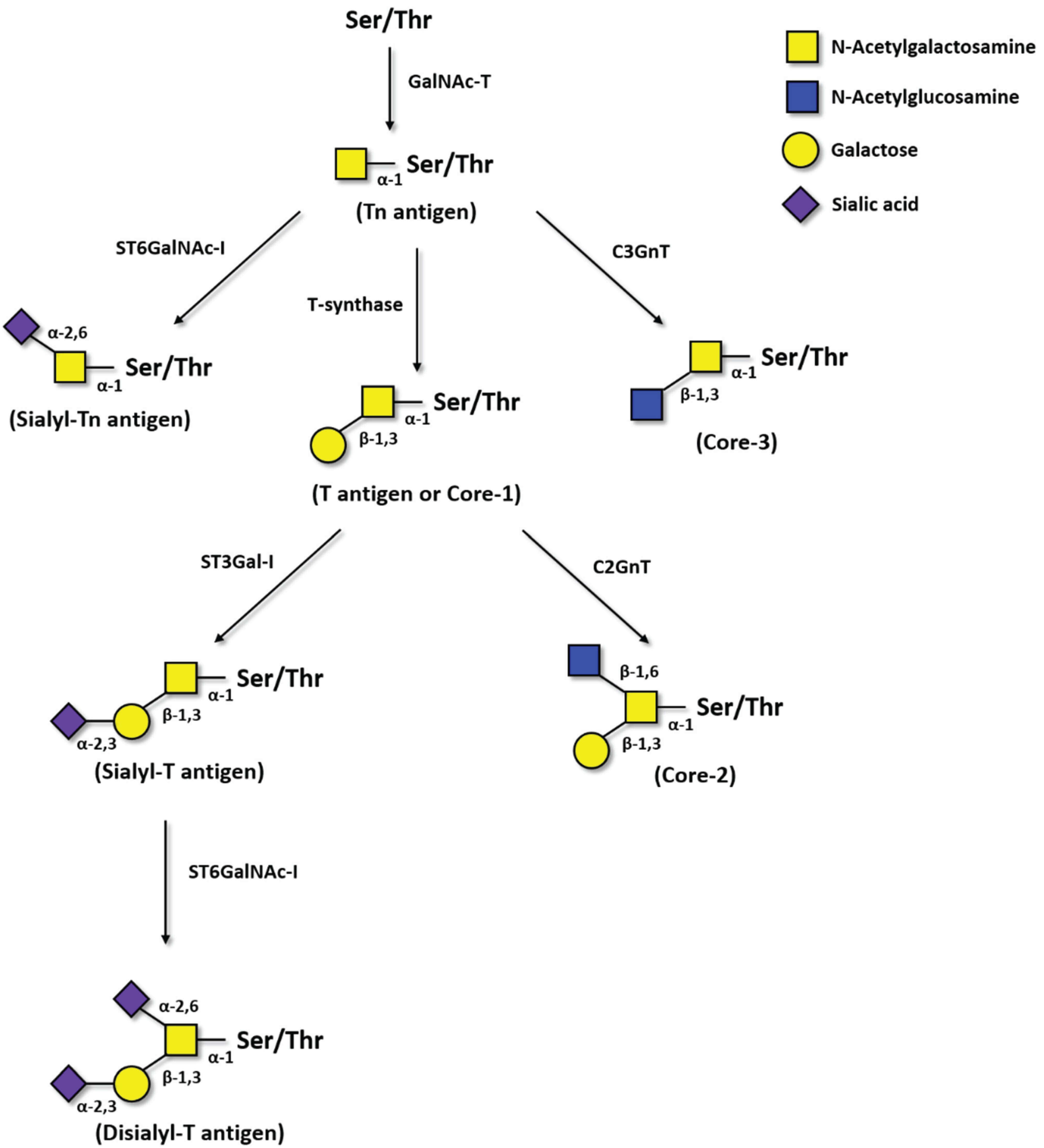 Fig.1 Flowchart of the biosynthesis pathways for Tn, T, sTn, and sT antigens.1
The T, sT, Tn, and sTn antigens are all related. They are all built during a process called O-glycosylation. The diagram below shows how this pathway works and how it becomes faulty in cancer. This entire process starts on a protein backbone, at a Serine (Ser) or Threonine (Thr) amino acid.
- Tn Formation: The very first step is an enzyme (GalNAc-T) attaching a single sugar, N-Acetylgalactosamine (GalNAc), to the protein. This simple, one-sugar structure is the Tn antigen. In healthy cells, this structure is almost instantly built upon; it is only an intermediate.
- Making T: In a normal pathway, an enzyme called T-synthase adds a second sugar, Galactose, to the Tn antigen. This creates the two-sugar structure known as the T antigen.
- Making sT: From the T antigen, a different enzyme (ST3Gal-I) can add a Sialic acid molecule. This creates the Sialyl-T (sT) antigen.
- Making sTn: If the T-synthase enzyme is missing or inactive, the cell cannot make the T antigen. Instead, a different enzyme (ST6GalNAc-I) adds a Sialic acid molecule directly to the original Tn antigen. This creates the Sialyl-Tn (sTn) antigen.
In cancer, this process is flawed. The enzymes that build longer chains (like T-synthase) are often suppressed. At the same time, the enzymes that add sialic acid molecules (such as ST6GalNAc-I and ST3Gal-I) are usually overactive. This combination leads to a massive accumulation of the short, truncated antigens: Tn, sTn, and sT.
Comparing the Key TACA Targets
Understanding the minor structural differences between these antigens is critical for cancer research. Each antigen provides different information about the cancer cell and requires a unique tool to study it.
| Antigen | Structure (Simple) | Expression in Healthy Tissue (Cancer Specificity) | Key Role in Cancer | Recommended Service |
|---|---|---|---|---|
| Tn Antigen |
Single Sugar (GalNAc-Ser/Thr) |
Very High Specificity. Usually, an internal precursor is not exposed on the surfaces of healthy cells. |
Proliferation, invasion, and adhesion. Its appearance is an early sign of malignancy. | Anti-Tn Antibody Development |
| sTn Antigen |
Capped Single Sugar (Sialic acid on Tn) |
Highest Specificity. Not a typical precursor. Its expression is a pathological event. |
Strongly associated with metastasis, poor prognosis, and immune evasion. | Anti-sTn Antibody Development |
| T Antigen |
Two Sugars (Gal-GalNAc-Ser/Thr) |
Low Specificity. Exposed on some healthy tissues, inflamed tissues, and red blood cells. |
Promotes cell-to-cell adhesion (via galectin binding) and proliferation. | Anti-T/sT Antibody Development |
| sT Antigen |
Capped Two Sugars (Sialic acid on T) |
Medium-Low Specificity. Found in some healthy tissues but highly overexpressed in cancer. |
Critical for adhesion to blood vessel walls (via selectin binding) and metastasis. | Anti-T/sT Antibody Development |
Why This Matters for Your Research
These differences are not just academic. The high cancer specificity of Tn and sTn makes them exceptional targets for developing new therapies, as an antibody against them is less likely to interact with healthy cells. In contrast, T and sT are vital for understanding the mechanisms of how cancer spreads. An sT antibody, for example, can be used to physically block a cancer cell from binding to a blood vessel. Because these structures are so similar, an antibody with poor specificity—one that accidentally binds both T and sT, or Tn and sTn—will produce confusing and unreliable data. A particular, custom-developed antibody from Creative Biolabs is the most critical tool for ensuring your adhesion studies are accurate and publishable.
Request a Quote for Your Project
Our Solution at Creative Biolabs: A True Custom Antibody Service
Creative Biolabs overcomes these challenges by combining expert glycobiology knowledge with advanced antibody engineering platforms. We do not offer a one-size-fits-all product. We offer a fully custom antibody service to build the precise tool you need. Our service is built on a foundation of precision, validation, and collaboration. We leverage multiple state-of-the-art platforms to guarantee the best possible outcome for your project.
This is our platform of choice for challenging glycan targets. Phage display is an in vitro method that uses phages to display billions of different human antibody fragments (scFv or Fab) on their surface. This creates a massive library of potential binders. We prepare highly purified, synthetic T antigen or sT antigen targets. We then screen the library against this target, fishing out only the phages that bind. This process is fast, highly controlled, produces recombinant antibodies (with known sequences), and avoids the use of animals. The true power of phage display is in negative selection. To create a highly specific T antibody, we first select for everything that binds T. Then, we take those binders and expose them to sT antigen. Any antibody that also binds sT is discarded. This leaves us with a pool of antibodies that are highly specific to T and do not see sT. We do the reverse to create a specific sT antibody.
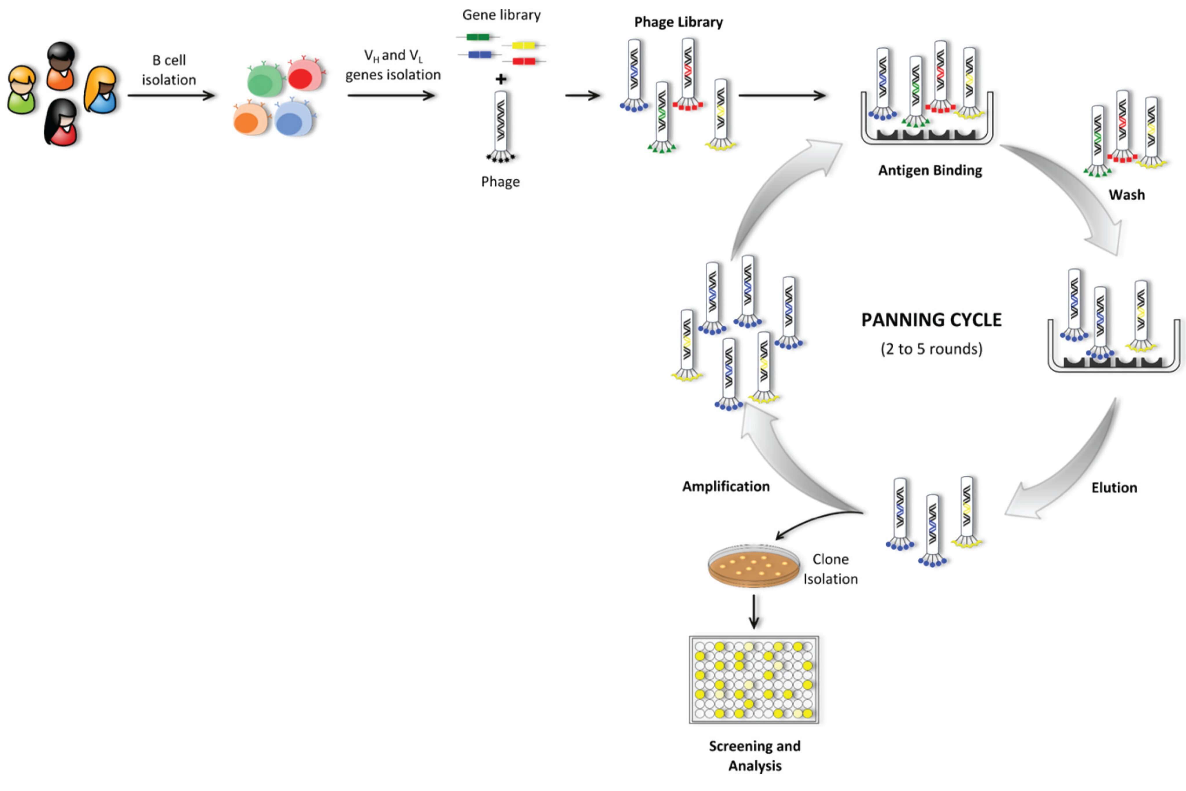 Fig.2 The phage display workflow.1
For researchers who prefer a traditional, full-length mouse or rabbit monoclonal antibody, we offer optimized hybridoma development. We conjugate the synthetic T or sT glycan to a large carrier protein to make it highly immunogenic. We then employ a rigorous screening process to identify hybridoma clones that produce antibodies with the exact specificity you require.
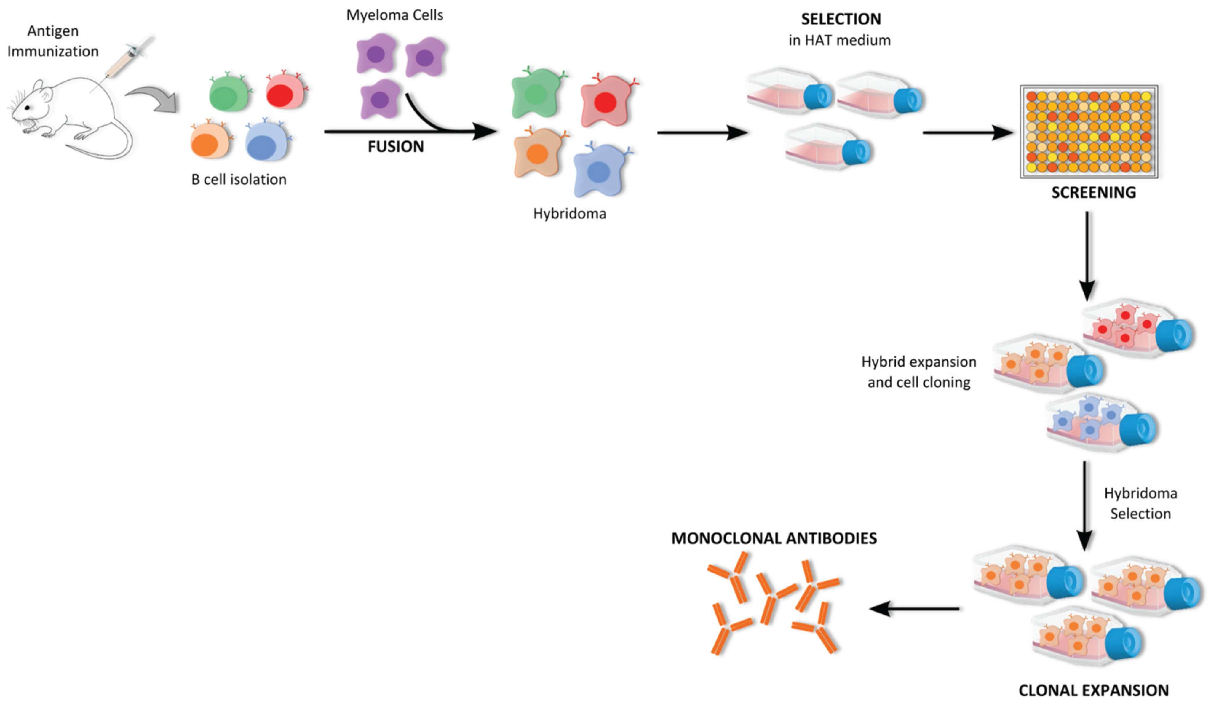 Fig.3 The hybridoma workflow.1
Your Project, Our Process: The Creative Biolabs Workflow
The T and sT antigens are at the heart of cancer cell adhesion and metastasis. Studying them effectively is a crucial step toward understanding and, ultimately, controlling the spread of cancer. But this research is impossible without tools that are precise, reliable, and specific. Stop relying on ambiguous data from non-specific antibodies. Let us build the exact tool you need. Contact our team at Creative Biolabs today for a free, no-obligation consultation on your project. Please tell us what you need to discover, and we will build the antibody to help you do it.
Reference:
- Loureiro, Liliana R., et al. "Challenges in antibody development against Tn and Sialyl-Tn antigens." Biomolecules 5.3 (2015): 1783-1809. Distributed under Open Access license CC BY 4.0, without modification. https://doi.org/10.3390/biom5031783
Supports
- TACAs Overview
- Guide to Blood Group Antigens
- Comparing sLeA and sLeX Roles in Cancer
- CA19-9 as a Pancreatic Cancer Biomarker
- Lewis Antigen System Overview
- TACA-Targeted ADCs, CAR-Ts, and RICs